. 2004 Jul;15(7):3083-94.
doi: 10.1091/mbc.e03-12-0888. Epub 2004 Apr 9.

Role of the midbody matrix in cytokinesis: RNAi and genetic rescue analysis of the mammalian motor protein CHO1

Affiliations

Role of the midbody matrix in cytokinesis: RNAi and genetic rescue analysis of the mammalian motor protein CHO1

Jurgita Matuliene et al. Mol Biol Cell. 2004 Jul.

Abstract

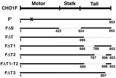
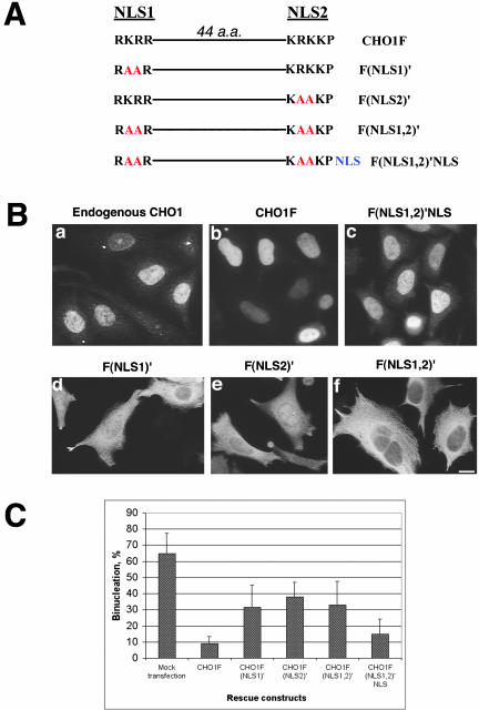
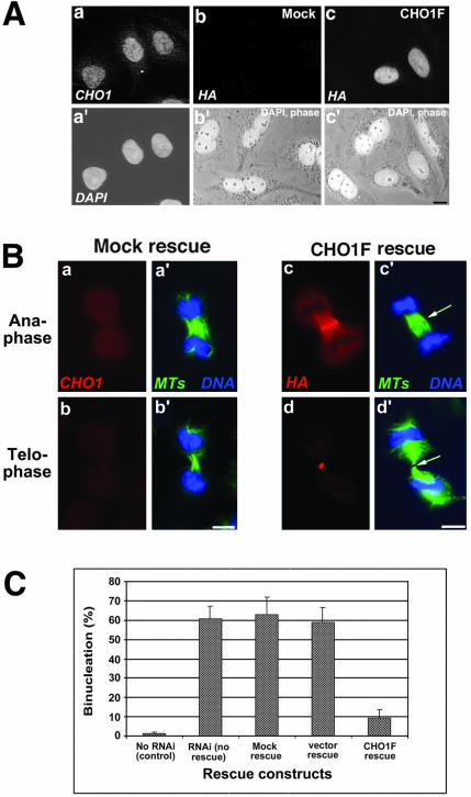
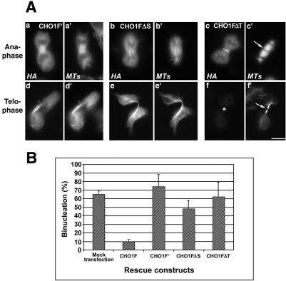
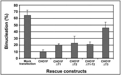
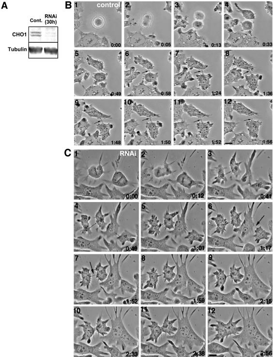
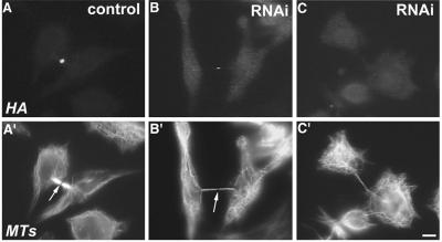
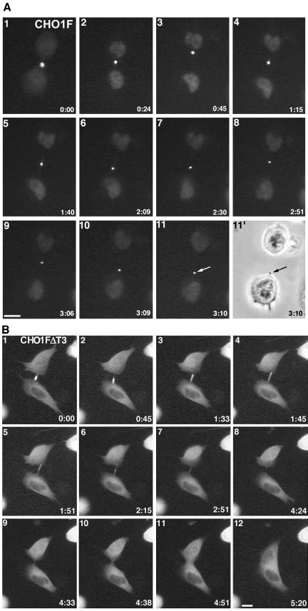
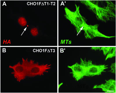
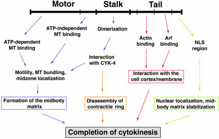
CHO1 is a kinesin-like motor protein essential for cytokinesis in mammalian cells. To analyze how CHO1 functions, we established RNAi and genetic rescue assays. CHO1-depleted cells reached a late stage of cytokinesis but fused back to form binucleate cells because of the absence of the midbody matrix in the middle of the intercellular bridge. Expression of exogenous CHO1 restored the formation of the midbody matrix and rescued cytokinesis in siRNA-treated cells. By analyzing phenotypes rescued with different constructs, it was shown that both motor and stalk domains function in midbody formation, whereas the tail is essential for completion of cytokinesis after the midbody matrix has formed. During the terminal stage of cytokinesis, different subregions of the tail play distinctive roles in stabilizing the midbody matrix and maintaining an association between the midbody and cell cortex. These results demonstrate that CHO1 consists of functionally differentiated subregions that act in concert to ensure complete cell separation.


Figures

Figure 1.
Figure 1.
A map of CHO1 constructs used for RNAi-rescue experiments. The number of amino acids, encoded by each construct, is identified beneath the bold lines. All constructs have four nucleotide changes in the region complementary to siRNA used for RNAi assays and are tagged with the HA epitope at the N terminus. X indicates the mutation in the ATP-binding site in the motor domain.
Figure 9.
Figure 9.
Nuclear localization is important for the function of CHO1 in cytokinesis. (A) Schematic representation of NLS-containing region in wild-type CHO1 and its NLS mutants. Amino acid changes within the sequence of the first NLS (NLS1), the second NLS (NLS2), and both NLS (NLS1,2) are shown in red. NLS shown in blue represents three SV40-derived nuclear localization signals fused to the C terminus of CHO1F(NLS1,2)′ construct to make CHO1F(NLS1,2)′NLS construct. (B) Endogenous CHO1 and exogenous CHO1F localize to the nucleus in interphase cells (a and b). Mutation in NLS1 and NLS2 significantly reduce the localization of CHO1 to the nucleus (d and e). Although nuclear localization is still detected in some cells expressing CHO1F(NLS1)′ and CHO1F(NLS2)′ constructs, cytoplasmic localization is more prominent in the majority of cells. Mutations introduced in both NLS completely inhibit nuclear targeting of CHO1 (f). Foreign NLS fused to the C terminus of CHO1F(NLS1,2)′ construct restores the CHO1 localization to the nucleus (c). (C) Rescue histogram shows a threefold increase in the level of binucleation after alteration of any one or both NLS. Frequency of binucleation is reduced twofold by fusing the viral NLS to the C terminus of CHO1F(NLS1,2)′ mutant, indicating the importance of nuclear localization for the function of CHO1 in cytokinesis. Cells were stained with polyclonal anti-CHO1 (B-a) and anti-HA antibody (B-b to f). Bar, 10 μm.
Figure 4.
Figure 4.
Exogenous CHO1F rescues cytokinesis in siRNA-transfected cells. (A) Exogenous CHO1F localizes to the nucleus similarly to endogenous CHO1 and reduces binucleation in siRNA-treated cells. a and a′: Localization of endogenous CHO1 seen by polyclonal anti-CHO1 antibody staining (a) and DAPI (a′). b and b′: Mock-rescued cells are not recognized by HA antibody (b) and are binucleate. c and c′: CHO1F-rescued cells are stained by monoclonal anti-HA antibody (c) and contain a single nucleus, in contrast to nonexpressing binucleate cells seen in the same field. (B) Exogenous CHO1F localizes to the midzone/midbody region (c) in an identical manner to endogenous CHO1 (unpublished data) and restores the organization of midzone and midbody matrix in siRNA-treated cells (arrows in c′ and d′). This is in contrast with mock-rescued cells, which still have midzones composed of disorganized microtubule arrays (a′ and b′). Cells are stained with polyclonal anti-CHO1 (a and b), polyclonal anti-HA (c and d), anti-α-tubulin (a′-d′; green) antibodies and DAPI (a′-d′; blue). Bars, 10 μm. (C) Histograms show the percentage of binucleate cells in control (no RNAi) cells, not rescued RNAi-affected cells, and cells rescued with mock, pCMV-HA vector alone, and CHO1F construct 30 h after transfection with siRNA. Binucleation in CHO1F-expressing cells is reduced sixfold in comparison with other types of rescue or no rescue at all.
Figure 5.
Figure 5.
ATP-binding mutant of CHO1 (CHO1F′) and CHO1 constructs lacking stalk (CHO1FΔS) or tail (CHO1FΔT) domains failed to rescue cytokinesis in endogenous CHO1-depleted cells. (A) CHO1F′ (a and d) and CHO1FΔS (b and e) constructs decorate the spindle microtubules throughout mitosis, but lack the ability to concentrate at the midzone/midbody region. In contrast to CHO1FΔT (c and f), CHO1F′ and CHO1FΔS constructs do not facilitate formation of the midbody matrix, based on continuous α-tubulin staining in the middle of the intercellular bridge (d′ and e′). CHO1FΔT localizes at the midzone (c) and midbody (f) and facilitates formation of the dense matrix (arrows in c′ and f′). Cells were stained with polyclonal anti-HA (a-f) and anti-α-tubulin (a′-f′) antibodies. Bar, 10 μm. (B) Histograms show no substantial decrease in the level of binucleation after rescue with CHO1F′, CHO1FΔS, and CHO1FΔT constructs.
Figure 6.
Figure 6.
CHO1 constructs lacking different regions of the tail domain show different abilities to rescue cytokinesis in endogenous CHO1-depleted cells. Histograms show the percentage of binucleation in rescued cells. CHO1 lacking T1 (CHO1FΔT1) or T2 (CHO1FΔT2), or both (CHO1FΔT1-T2) are half as efficient in the rescue of cytokinesis, whereas the construct lacking NLS region (CHO1FΔT3) is one quarter as efficient in comparison to the wild-type CHO1F.
Figure 2.
Figure 2.
RNAi against CHO1 causes depletion of CHO1 protein and inhibition of cytokinesis in CHO cells. (A) Cells were transfected with siRNA (RNAi) or transfection reagent alone (control) and analyzed by immunoblotting for the presence of CHO1 protein 30 h after transfection. Immunoblotting was performed using monoclonal anti-CHO1 and anti-α-tubulin antibodies for the loading control. CHO1 antibodies reveal two isoforms, MKLP1 and CHO1, coexpressed in a single cell (Kuriyama et al., 2002). (B) Successful division of mock-transfected cells monitored by time-lapse phase-contrast microscopy. A dark midbody dot indicated by arrows is seen in the middle of the intercellular bridge until complete separation of the daughter cells. (C) Time-lapse phase-contrast microscopy of RNAi-affected cells. Cells reach the late stage of cytokinesis, but fuse back within 1-2 h after formation of the intercellular bridge in which the dark midbody structures are generally missing (arrows). Bars, 10 μm.
Figure 3.
Figure 3.
RNAi against CHO1 affects the thickness and the length of intercellular bridges and the formation of midbody matrix in dividing cells. Control cell (A) and siRNA-transfected cells (B and C) were fixed at the late stage of cell division and stained with polyclonal anti-CHO1 (A-C) and anti-α-tubulin (A′-C′) antibodies. The control daughter cells are connected by a thick intercellular bridge with a well-defined midbody matrix, which cannot be penetrated by anti-α-tubulin antibody and, therefore, appears as a dark region (arrow in A′). When CHO1 is depleted from the midbody region (B and C), the intercellular bridges become longer and thinner (B′ and C′), and the midbody matrix is faint (arrow in B′) or hardly detectable in the cells with the lowest amounts of CHO1 expression (C′). Bar, 10 μm.
Figure 7.
Figure 7.
The NLS-containing region of the CHO1 tail is required for stabilization of the midbody matrix. (A) The midbody is stable in a GFP-CHO1F-rescued cell monitored by the time-lapse fluorescence microscopy. At the end of cytokinesis, the intercellular bridge is pinched off at one side of the midbody, and the midbody is incorporated into one of the daughter cells (arrows). (B) Time-lapse fluorescence video and assembled micrographs shows that the midbody in a GFP-CHO1FΔT3-rescued cell gradually disintegrates, causing almost completely separated two daughter cells to merge again. Note that full-length CHO1F localizes to the nucleus after the nuclear envelope is formed (frames 2-11 in 7A), whereas the construct-lacking NLS region remains in the cytoplasm throughout interphase (B). Bars, 10 μm.
Figure 8.
Figure 8.
Structural stability of the midbody/midbody matrix formed in cells expressing CHO1FΔT1-T2 (A and A′) and CHO1FΔT3 (B and B′). Cells were double-stained with anti-HA (A and B) and anti-α-tubulin antibodies (B and B′). Although the midbody remains inside the fused cells rescued with CHO1FΔT1-T2 construct, the CHO1FΔT3-containing midbody disintegrates. Bars, 10 μm.
Figure 10.
Figure 10.
A model of CHO1 functions in cytokinesis.

References

    1. Abaza, A., Soleilhac, J.M., Westendorf, J., Piel, M. Crevel, I., Roux, A., and Pirollet, F. (2003). M phase phosphoprotein 1 is a human plus-end-directed kinesin-related protein required for cytokinesis. J. Biol. Chem. 278, 27844-27852. - PMC - PubMed
    1. Adams, R.R., Tavares, A.A.M., Salzberg, A., Bellen, H.J., and Glover, D.M. (1998). pavarotti encodes a kinesin-like protein required to organize the central spindle and contractile ring for cytokinesis. Genes Dev. 12, 1483-1494. - PMC - PubMed
    1. Alsop, G.B., and Zhang, D. (2003). Microtubules are the only structural constituent of the spindle apparatus required for induction of cell cleavage. J. Cell Biol. 162, 383-390. - PMC - PubMed
    1. Boman, A.L., and Kahn, R.A. (1995). Arf proteins: the membrane traffic police? Trends Biochem. Sci. 20, 147-150. - PubMed
    1. Boman, A.L., J. Kuai, J., Zhu, X., Chen, J., Kuriyama, R., and Kahn, R.A. (1999). Arf proteins bind to mitotic kinesin-like protein 1 (MKLP1) in a GTP-dependent fashion. Cell Motil. Cytoskel. 44, 119-132. - PubMed

Publication types

MeSH terms