. 2004 Jun;15(6):2794-803.
doi: 10.1091/mbc.e03-12-0879. Epub 2004 Apr 9.

Src SH3/2 domain-mediated peripheral accumulation of Src and phospho-myosin is linked to deregulation of E-cadherin and the epithelial-mesenchymal transition

Affiliations

Src SH3/2 domain-mediated peripheral accumulation of Src and phospho-myosin is linked to deregulation of E-cadherin and the epithelial-mesenchymal transition

Egle Avizienyte et al. Mol Biol Cell. 2004 Jun.

Abstract

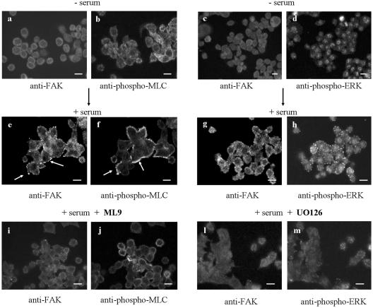
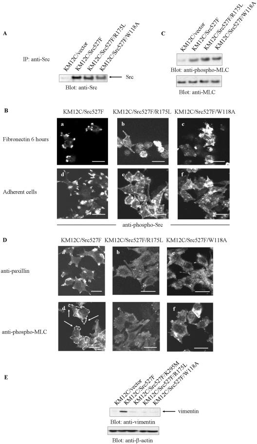
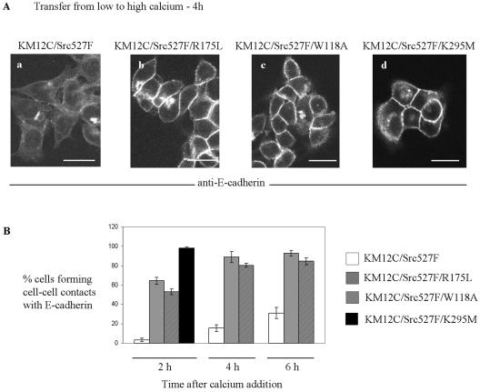
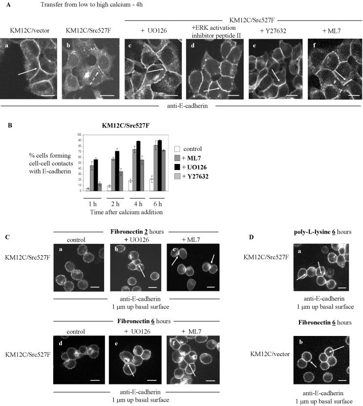
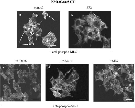
Elevated Src kinase in epithelial cancer cells induces adhesion changes that are associated with a mesenchymal-like state. We recently showed that Src induces dynamic integrin adhesions in KM12C colon cancer cells, whereas E-cadherin-dependent cell-cell contacts become disorganized. This promotes a fibroblastic-like morphology and expression of the mesenchymal marker vimentin. Furthermore, Src-induced deregulation of E-cadherin, and the associated mesenchymal transition, is dependent on integrin signaling (Avizienyte et al., Nat. Cell Biol. 2002, 4, 632-638), although the nature of downstream signals that mediate these Src- and integrin-dependent effects are unknown. Here we show that the SH2 and SH3 domains of Src mediate peripheral accumulation of phospho-myosin, leading to integrin adhesion complex assembly, whereas loss of SH2 or SH3 function restores normal regulation of E-cadherin and inhibits vimentin expression. Inhibitors of MEK, ROCK, or MLCK also suppress peripheral accumulation of phospho-myosin and Src-induced formation of integrin-dependent adhesions, whereas at the same time restoring E-cadherin redistribution to regions of cell-cell contact. Our data therefore implicate peripheral phospho-myosin activity as a point of convergence for upstream signals that regulate integrin- and E-cadherin-mediated adhesions. This further implicates spatially regulated contractile force as a determinant of epithelial cell plasticity, particularly in cancer cells that can switch between epithelial and mesenchymal-like states.


Figures

Figure 1.
Figure 1.
ERK/MLCK activities are required for Src-induced integrin adhesion assembly in KM12C colon cancer cells. (A) KM12C cells transfected with Src527F (KM12C/Src527F) were cultured in uncoated plastic dishes (adherent) or in poly-HEMA–coated dishes (suspension). Phospho-ERK or phospho-MLC levels were detected by probing total lysates with the anti-phospho-ERK (Thr202/Tyr204) or anti-phospho-MLC (Ser19) antibodies (top panels). The filters were reprobed with anti-ERK or anti-MLC antibodies (lower panels). (B) Increased level of total cellular phospho-MLC in KM12C/Src527F cells was blocked by MLCK inhibitor ML9 (7.6 μM). (C) (a–f) KM12C/Src527F (a–c) or KM12C/vector (d–f) cells were plated on fibronectin-coated substratum for 6 h, fixed, and stained with anti-paxillin (a and d), anti-phospho-ERK (Thr202/Tyr204; b and e) or anti-phospho-MLC (Ser19; c and f) antibodies. Arrows show paxillin, phospho-ERK and phospho-MLC localized at cell-matrix adhesion complexes at the ends of protrusive structures in KM12C/Src527F cells. (g–m) MEK inhibitor UO126 (25 μM; g–i) or MLCK inhibitor ML7 (5 μM; j–m) blocked the formation of protrusive integrin-mediated adhesions in KM12C/Src527F cells. Arrows show localization of paxillin or phospho-ERK at nonprotrusive cell-matrix adhesion structures at the cell periphery. Scale bars, 10 μm. (D) KM12C/Src527F cells were plated on poly-l-lysine–coated substratum, fixed, and stained with anti-paxillin (a), anti-phospho-ERK (Thr202/Tyr204; b) or anti-phospho-MLC (Ser19; c) antibodies. Scale bars, 10 μm.
Figure 2.
Figure 2.
Serum induces peripheral integrin adhesion structures in KM12C cells via MEK/ERK and MLCK activities. (a--d) KM12C/vector cells were switched to serum-free low-calcium medium (KGM) for 24 h, fixed, and costained with anti-FAK and anti-phospho-MLC (Ser19; a and b) or anti-FAK and anti-phospho-ERK (Thr202/Tyr204) (c and d) antibodies. Scale bars, 10 μm. (e–h) Serum-starved KM12C/vector cells were stimulated with MEM containing 10% FBS for 10 min, fixed, and costained with anti-FAK and anti-phospho-MLC (Ser19) (e and f) or anti-FAK and anti-phospho-ERK (Thr202/Tyr204) antibodies (g and h). Arrows in e indicate the formation of peripheral focal adhesion complexes after stimulation with serum. Arrows in f show serum-induced accumulation of phospho-MLC at the cell periphery. Scale bars, 10 μm. (i–m) KM12C/vector cells were treated as described above except that stimulation with serum was carried out in the presence of ML9 (7.6 μM; i and j) or UO126 (25 μM; l and m). Cells were fixed and costained with anti-FAK and anti-phospho-MLC (Ser19) (i and j) or anti-FAK and anti-phospho-ERK (Thr202/Tyr204) antibodies (l and m). Scale bars, 10 μm.
Figure 3.
Figure 3.
SH2 and SH3 domains of Src are required for accumulation of phospho-MLC at the cell periphery. (A) Expression levels of mutant Src kinase proteins in KM12C cells. (B) KM12C/Src527F, KM12C/Src527F/R175L, or KM12C/Src527F/W118A cells were plated on fibronectin for 6 h (a–c) or cultured in MEM and switched to low-calcium medium (d–f). Cells were fixed and stained with anti-phospho-Src (Tyr416) antibody. Scale bars, 20 μm. (C) Phospho-MLC levels in KM12C/vector, KM12C/Src527F, KM12C/Src527F/R175L, or KM12C/Src527F/W118A cells were detected by probing total lysates with the anti-phospho-MLC (Ser19) antibody (top panel). The filters were reprobed with anti-MLC antibody (bottom panel). (D) KM12C/Src527F, KM12C/Src527F/R175L, or KM12C/Src527F/W118A cells were switched to low-calcium medium, fixed, and stained with anti-paxillin (a–c) or anti-phospho-MLC (Ser19) (d–f) antibody. Arrows in d show accumulation of phospho-MLC at the cell periphery. Scale bars, 20 μm. (E) Vimentin expression levels in KM12C/vector, KM12C/Src527F, KM12C/Src527F/K295M, KM12C/Src527F/R175L, or KM12C/Src527F/W118A cells.
Figure 4.
Figure 4.
Src kinase activity at peripheral integrin-mediated adhesion complexes is required to impair E-cadherin localization. (A) KM12C cells expressing Src527F (a), Src527F/R175L (b), Src527F/W118A (c) or kinase-defective Src527F/K295M) (d) were switched from low- to high-calcium medium for 4 h. Cells were fixed and stained with anti-E-cadherin. Scale bars, 20 μm. (B) Quantitation of percentage of cells which localize E-cadherin to cell-cell contacts after switch to high-calcium medium. The mean and range of two independent experiments is shown.
Figure 6.
Figure 6.
Inhibition of MEK/ERK, ROCK, and MLCK activities reverses Src-induced deregulation of E-cadherin–associated cell-cell contacts. (A) KM12C/vector or KM12C/Src527F cells were switched from low- to high-calcium medium for 4 h (a and b). MEK inhibitor UO126 (25 μM; c), ERK activation inhibitor peptide II (100 μM; d), ROCK inhibitor Y27632 (10 μm; e), or MLCK inhibitor ML7 (5 μM; f) was added to high-calcium medium and KM12C/Src527F cells were maintained for 4 h in such medium. Cells were fixed and stained with anti–E-cadherin antibody. Solid arrows show accumulation of E-cadherin at cell-cell contacts in KM12C/vector cells and KM12C/Src527F cells treated with either UO126, ERK activation inhibitor peptide II, Y27632, or ML7 (a, c, and d–f). Broken arrow in b points to disrupted E-cadherin staining in KM12C/Src527F cells after the switch to high-calcium medium. Scale bars, 10 μm. (B) Quantitation of percentage of KM12C/Src527F cells that are forming cadherin-mediated cell-cell contacts when cells are switched to high-calcium medium containing ML7, UO126, or Y27632. (C) KM12C/Src527F were plated on fibronectin-coated substratum for 2 or6h(a–f) without inhibitors (a and d) or with the MEK inhibitor UO126 (25 μM; b and e) or MLCK inhibitor ML7 (5 μM; c and f). Cells were fixed and stained with anti–E-cadherin antibody. Arrows show accumulation of E-cadherin at cell-cell contacts in KM12C/Src527F cells plated on fibronectin when MEK/ERK or MLCK activity is inhibited. Scale bars, 10 μm. (D) (a) KM12C/Src527F were plated on poly-l-lysine–coated substratum for 6 h and E-cadherin was visualized by staining with anti–E-cadherin antibody. Arrows indicate accumulation of E-cadherin between active Src527F expressing cells when integrin signaling is suppressed. (b) KM12C/vector cells were plated on fibronectin for 6 h, fixed, and stained with anti–E-cadherin antibody. Arrows show accumulation of E-cadherin at cell-cell contacts. Scale bars, 10 μm.
Figure 5.
Figure 5.
Inhibition of Src kinase, MEK/ERK, ROCK, or MLCK activity impairs localization of phospho-MLC at the cell periphery. KM12C/Src527F cells were cultured in high-calcium medium (MEM), further maintained in low-calcium medium and treated with either PP2 (20 μM; b), UO126 (25 μM; c), Y27632 (10 μM; d), or ML7 (5 μM; e) for 4 h. Cells were fixed and stained with anti-phospho-MLC (Ser19) antibody. Arrows show phospho-MLC localization at the leading edge of protrusive adhesion structures. Scale bars, 10 μm.
Figure 7.
Figure 7.
Inhibition of PI 3-kinase or STAT3 activity does not induce cell-cell contact formation in active Src-expressing cells. (A) KM12C cells expressing active Src527F were treated with the PI 3-kinase inhibitor LY294002 (50 μM). Cell lysates were probed with anti-phospho-Akt (Ser473) or anti-Akt antibody (left panels). Levels of active STAT3 (phospho-Tyr705) and total STAT3 in KM12C/Src527F cells or A431 cells stimulated with EGF are shown in the right panels. (B) Percentage of KM12C/Src527F cells that are forming cadherin-mediated cell-cell contacts after the cells are switched to high-calcium medium without inhibitors or with ERK activation inhibitory peptide II (100 μM), STAT3 inhibitory peptide (100 μM), or PI 3-kinase inhibitor LY294002 (50 μM).

References

    1. Avizienyte, E., Wyke, A.W., Jones, R.J., McLean, G.W., Westhoff, M.A., Brunton, V.G., and Frame, M.C. (2002). Src-induced de-regulation of E-cadherin in colon cancer cells requires integrin signalling. Nat. Cell Biol. 4, 632-638. - PubMed
    1. Bibbins, K.B., Boeuf, H., and Varmus, H.E. (1993). Binding of the Src SH2 domain to phosphopeptides is determined by residues in both the SH2 domain and the phosphopeptides. Mol. Cell. Biol. 13, 7278-7287. - PMC - PubMed
    1. Bowman, T. et al. (2001). Stat3-mediated Myc expression is required for Src transformation and PDGF-induced mitogenesis. Proc. Natl. Acad. Sci. USA 98, 7319-7324. - PMC - PubMed
    1. Boyer, B., Roche, S., Denoyelle, M., and Thiery, J.P. (1997). Src and Ras are involved in separate pathways in epithelial cell scattering. EMBO J. 16, 5904-5913. - PMC - PubMed
    1. Boyer, B., Bourgeois, Y., and Poupon, M.F. (2002). Src kinase contributes to the metastatic spread of carcinoma cells. Oncogene 21, 2347-2356. - PubMed

Publication types

MeSH terms

LinkOut - more resources