. 2004 Apr 20;101(16):6027-32.
doi: 10.1073/pnas.0401367101. Epub 2004 Apr 9.

BMP4 supports self-renewal of embryonic stem cells by inhibiting mitogen-activated protein kinase pathways

Affiliations

BMP4 supports self-renewal of embryonic stem cells by inhibiting mitogen-activated protein kinase pathways

Xiaoxia Qi et al. Proc Natl Acad Sci U S A. .

Abstract

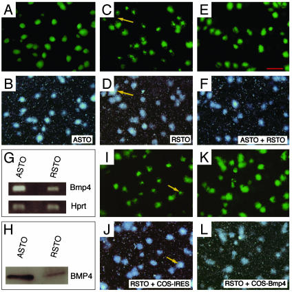
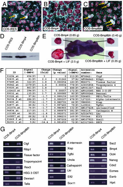
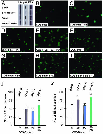
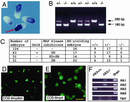
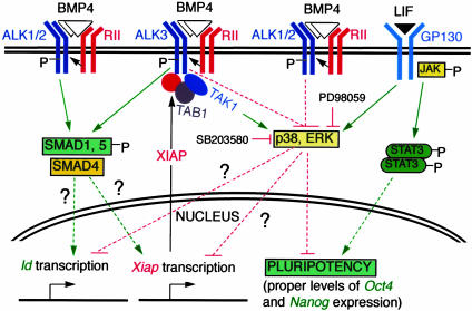
The fate of pluripotent stem cells is tightly controlled during early embryonic development. Both the derivation and the maintenance of embryonic stem cells (ES cells) in vitro depend on feeder cell-derived growth factors that are largely unidentified. To dissect the mechanisms governing pluripotency, we conducted a screen to identify factors that are produced by mouse embryonic fibroblast STO cells and are required to maintain the pluripotency of ES cells. One of the factors is bone morphogenetic protein 4 (BMP4). Unexpectedly, the major effect of BMP4 on the self-renewal of ES cells is accomplished by means of the inhibition of both extracellular receptor kinase (ERK) and p38 mitogen-activated protein kinase (MAPK) pathways, and inhibitors of ERK and p38 MAPKs mimic the effect of BMP4 on ES cells. Importantly, inhibition of the p38 MAPK pathway by SB203580 overcomes the block in deriving ES cells from blastocysts lacking a functional Alk3, the BMP type IA receptor. These results uncover a paradigm for BMP signaling in the biology of pluripotent stem cells.


Figures

Fig. 1.
Fig. 1.
Identification of BMP4 as one of the STO-produced factors mimicking ASTO activity. We plated ≈2,000 ES cells onto a 35-mm dish containing ASTO or RSTO cells. Photos were taken 5 days after the culture with complete ES cell medium. (A and B) Fluorescent and dark-field photomicrographs of ES cell colonies on ASTO. All colonies on ASTO had robust Oct4-GFP expression. (C and D) Fluorescent and dark-field photomicrographs of ES cell colonies on RSTO. Partial loss of GFP was obvious in many colonies, and a big colony only expressed GFP in a small area (arrow). (E and F) ES cells cultured on an equal mixture of ASTO and RSTO. The Oct4-GFP expression resembled those in A and B. (G) RT-PCR showing ASTO expressed higher levels of Bmp4 than RSTO with Hprt RT-PCR for normalization. (H) Western blotting showing ASTO-conditioned medium containing higher levels of BMP4 than RSTO-conditioned medium (equal loading). (I and J) ES cells cultured on RSTO with COS-IRES-conditioned medium had a similar phenotype as those shown in C and D. (K and L) ES cells cultured on RSTO with COS-Bmp4-conditioned medium showed robust Oct4-GFP expression in almost all colonies. Scale bar in AF and IL, 700 μm.
Fig. 2.
Fig. 2.
BMP4 prevents the differentiation of ES cells. (AC) We plated ≈2 × 10⁵ ES cells onto each 35-mm dish with COS-IRES, COS-Bmp4, or COS-Bmp8bh as feeders. After culture for 5 days with complete ES cell medium, cells were fixed in 4% paraformaldehyde/PBS and stained for ALP. (A) ES cells grown on COS-IRES. Loss of ALP was observed in some colonies (arrows). (B) ES cells grown on COS-Bmp4. Little loss of ALP was observed. (C) ES cells grown on COS-Bmp8bh. More colonies lost ALP expression than in A. (D) Western blotting to detect BMP4 proteins in conditioned medium from COS cells. COS-IRES-conditioned medium contained BMP4 corresponding to the endogenous expression. COS-Bmp4 expressed BMP4 at higher levels, whereas COS-Bmp8bh-conditioned medium only contained very low levels of BMP4. (E) Teratoma formation assay. ES cells were cultured on COS-Bmp4 and COS-Bmp8bh with or without LIF for 3 days. We injected ≈2 × 10⁶ ES cells in 50 μl of PBS from these cultures into 129/SvEv males s.c.. Tumors were observed and dissected 4 weeks after injection, with tumor weight labeled for each culture condition. (F) Shown are 17 differentially expressed sequences between ES cells cultured on COS-Bmp4 and COS-Bmp8bh (P < 0.015). (G) Semiquantitative RT-PCR to confirm 17 differentially expressed sequences. Except Ssc2 (no difference), all sequences were shown to be consistent with the microarray data shown in F. RT-PCR also showed that Bmp4, Oct4, and Nanog were expressed in both groups of ES cells at similar levels. After 45 cycles of amplification, markers for trophoblast Cdx2, Eomes, and Esrrb were detected without significant differences between the two samples. Scale bar in AC, 700 μm.
Fig. 3.
Fig. 3.
Relationship of BMP4 signaling and MAPK pathways. (A) BMP4 inhibits p38 and ERK MAPKs. A Bmp4-/- ES cell line was used for these studies. The levels of phospho-p38 and ERK were reduced significantly 5 min after BMP4 addition (50 ng/ml). A complete recovery for ERK and partial recovery for p38 were observed at 60 min. α-Tubulin was used as a loading control. (B) ES cells cultured on COS-IRES. We plated ≈5 × 10⁴ ES cells on a 35-mm dish that contained COS-IRES feeder cells. ES cells were cultured for 3 days and then photos were taken to show colonies. (C) ES cells cultured on COS-IRES with p38 inhibitor SB203580 (1 μM). Colonies were larger than those in B. (D) ES cells cultured on COS-IRES with MEK inhibitor PD98059 (12.5 μM). Colonies were similar to those in C.(E) ES cells cultured on COS-IRES with both PD98059 and SB203580. (F) ES cells cultured on COS-Bmp4. (G) ES cells cultured on COS-Bmp4 plus SB203580. (H) ES cells cultured on COS-Bmp4 with PD98059. (I) ES cells cultured on COS-Bmp4 plus both inhibitors. (J) Effects of MAPK inhibitors on colony formation of ES cells. We plated 500 ES cells on COS-Bmp8bh feeder cells in each of the 35-mm dish and cultured in ES cell medium with or without MAPK inhibitors. Colonies were counted 5 days later. Each group was in triplicate. (K) Colony formation experiments as described for J were conducted with COS-Bmp4 as feeders. *, P < 0.01. Scale bar in BI, 700 μm.
Fig. 4.
Fig. 4.
Derivation of Alk3-/- ES cells by inhibition of p38 MAPK. (A) Wholemount in situ hybridization of Alk3 mutant embryos at E6.0–6.5 with Oct4 antisense RNA probes. Only one embryo was Alk3-/- and showed a much higher level of Oct4 expression and wrinkled edges of the epiblast (arrow). The rest of the embryos were either wild type or Alk3+/-.(B) Genotype results for ES cells containing the Alk3 null allele in C.(C) Inhibition of p38 MAPK pathway rescued Alk3-/- ES cells. Culturing 124 blastocysts from Alk3+/- crosses in complete ES cell medium only yielded ES cells from wild type (n = 9) or heterozygotes (n = 17). With ERK pathway inhibitor PD98059 and G418 (180 μg/ml) in culture medium, 17 of 43 blastocysts gave rise to ES cells and all were Alk3+/-. With a combination of PD98059 and SB203580 in the medium, the following 14 of 31 blastocysts yielded ES cells: three wild type, nine heterozygotes, and two homozygotes. To test whether inhibition of p38 pathway alone was sufficient or not to derive Alk3-/- ES cells, 31 blastocysts were cultured in the presence of SB203580. Among 22 ES-cell-yielding blastocysts, 3 were Alk3-/- (4 wild-type and 15 heterozygotes). (D) Alk3-/- ES cells cultured on COS-Bmp8bh. We plated ≈1 × 10⁴ ES cells onto COS-Bmp8bh and cultured them in ES cell medium for 4 days. ES cell colonies were rather small. (E) Alk3-/- ES cells cultured on COS-Bmp4. ES cell colonies were much larger than those shown in D, indicating that Alk3-/- ES cells are still responsive to BMP4 and other BMP type I receptors compensate for the loss of Alk3. (F) Expression of putative BMP type I receptors in wild-type and Alk3-/- ES cells by RT-PCR. Brain cDNA was used as a control. Alk3 was the major type I BMP receptor in wild-type ES cells, whereas both Alk1 and Alk2 were present in Alk3-/- mutant ES cells. Scale bar in A, D, and E, 700 μm.
Fig. 5.
Fig. 5.
Schematic representation of BMP4 and LIF signaling in pluripotency. Upon dimeric BMP4 binding to the receptor complex, type II BMP receptor (RII) phosphorylates and activates type IA receptor (ALK3). Activated ALK3 causes the inactivation (dephosphorylation) of p38 and ERK MAPKs by unknown mechanisms. This inactivation relieves the negative effects of MAPKs on pluripotency (the proper levels of Oct4 and Nanog expression), as confirmed further by using MAPK inhibitors SB203580 and PD98059. As a result of the activation of ALK3, transcription of Xiap is up-regulated either by means of the inhibition of p38/ERK or activation of SMAD1/5/8 pathway. XIAP protein in the cytoplasm interacts with ALK3 and TAB1 to result in the activation of TAK1 by ALK3, consequently, a recovery of p38 activity. In the absence of ALK3, ALK1, or ALK2 proteins act as BMP4 type I receptor to maintain the pluripotency of ES cells. XIAP does not cause the loss of pluripotency in the absence of ALK3, presumably because of its inability to interact with ALK1 or ALK2. Lastly, the synergistic effect of BMP4 and LIF is likely attributed to the ability of BMP4 to antagonize the stimulation of ERK by LIF, inhibit p38 MAPK activity, and up-regulate Id genes and to the ability of LIF to activate JAK-STAT pathway. Solid lines indicate a direct effect.

References

    1. Martin, G. R. (1981) Proc. Natl. Acad. Sci. USA 78, 7634-7638. - PMC - PubMed
    1. Evans, M. J. & Kaufman, M. H. (1981) Nature 292, 154-156. - PubMed
    1. Bradley, A., Evans, M., Kaufman, M. H. & Robertson, E. (1984) Nature 309, 255-256. - PubMed
    1. Nichols, J., Zevnik, B., Anastassiadis, K., Niwa, H., Klewe-Nebenius, D., Chambers, I., Scholer, H. & Smith, A. (1998) Cell 95, 379-391. - PubMed
    1. Hanna, L. A., Foreman, R. K., Tarasenko, I. A., Kessler, D. S. & Labosky, P. A. (2002) Genes Dev. 16, 2650-2661. - PMC - PubMed

Publication types