

Identification and analysis of polyserine linker domains in prokaryotic proteins with emphasis on the marine bacterium Microbulbifer degradans
- PMID: 15075401
- PMCID: PMC2286767
- DOI: 10.1110/ps.03511604
Identification and analysis of polyserine linker domains in prokaryotic proteins with emphasis on the marine bacterium Microbulbifer degradans
Abstract
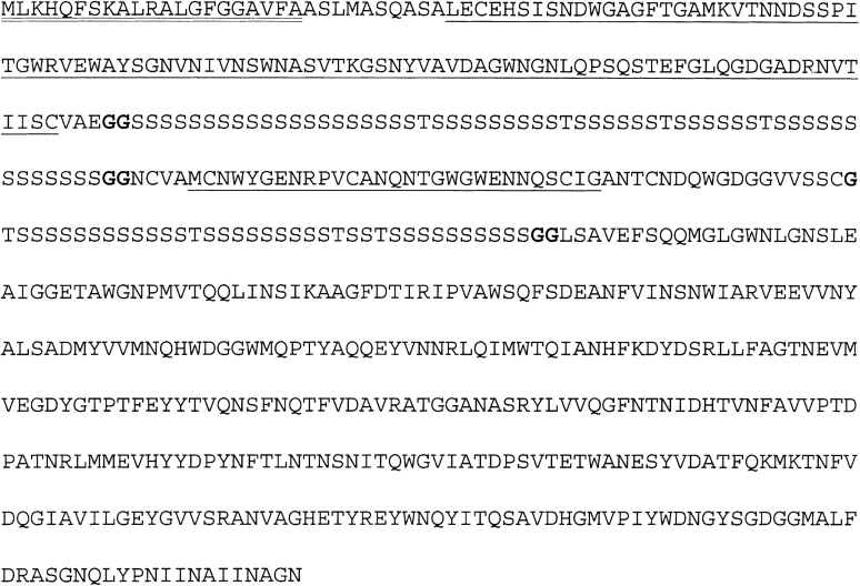
Polyserine linkers (PSLs) are interdomain, serine-rich sequences found in modular proteins. Though common among eukaryotes, their presence in prokaryotic enzymes is limited. We identified 46 extracellular proteins involved in complex carbohydrate degradation from Microbulbifer degradans that contain PSLs that separate carbohydrate-binding domains or catalytic domains from other binding domains. In nine M. degradans proteins, PSLs also separated amino-terminal lipoprotein acylation sites from the remainder of the polypeptide. Furthermore, among the 76 PSL proteins identified in sequence repositories, 65 are annotated as proteins involved in complex carbohydrate degradation. We discuss the notion that PSLs are flexible, disordered spacer regions that enhance substrate accessibility.
Figures
Similar articles
-
Genomic analysis and initial characterization of the chitinolytic system of Microbulbifer degradans strain 2-40.J Bacteriol. 2003 Jun;185(11):3352-60. doi: 10.1128/JB.185.11.3352-3360.2003. J Bacteriol. 2003. PMID: 12754233 Free PMC article.
-
Genomic and proteomic analyses of the agarolytic system expressed by Saccharophagus degradans 2-40.Appl Environ Microbiol. 2006 May;72(5):3396-405. doi: 10.1128/AEM.72.5.3396-3405.2006. Appl Environ Microbiol. 2006. PMID: 16672483 Free PMC article.
-
Feasibility test of utilizing Saccharophagus degradans 2-40(T) as the source of crude enzyme for the saccharification of lignocellulose.Bioprocess Biosyst Eng. 2014 Apr;37(4):707-10. doi: 10.1007/s00449-013-1040-1. Epub 2013 Aug 30. Bioprocess Biosyst Eng. 2014. PMID: 23990129
-
Complete genome sequence of the complex carbohydrate-degrading marine bacterium, Saccharophagus degradans strain 2-40 T.PLoS Genet. 2008 May 30;4(5):e1000087. doi: 10.1371/journal.pgen.1000087. PLoS Genet. 2008. PMID: 18516288 Free PMC article.
-
Carbohydrase systems of Saccharophagus degradans degrading marine complex polysaccharides.Mar Drugs. 2011;9(4):645-665. doi: 10.3390/md9040645. Epub 2011 Apr 21. Mar Drugs. 2011. PMID: 21731555 Free PMC article. Review.
Cited by
-
Structure-function analysis of the extracellular domain of the pneumococcal cell division site positioning protein MapZ.Nat Commun. 2016 Jun 27;7:12071. doi: 10.1038/ncomms12071. Nat Commun. 2016. PMID: 27346279 Free PMC article.
-
Mitochondrial selfish elements and the evolution of biological novelties.Curr Zool. 2016 Dec;62(6):687-697. doi: 10.1093/cz/zow044. Epub 2016 Mar 25. Curr Zool. 2016. PMID: 29491956 Free PMC article.
-
CelAB, a multifunctional cellulase encoded by Teredinibacter turnerae T7902T, a culturable symbiont isolated from the wood-boring marine bivalve Lyrodus pedicellatus.Appl Environ Microbiol. 2007 Dec;73(23):7785-8. doi: 10.1128/AEM.00876-07. Epub 2007 Oct 12. Appl Environ Microbiol. 2007. PMID: 17933945 Free PMC article.
-
Genome sequence of Streptococcus gallolyticus: insights into its adaptation to the bovine rumen and its ability to cause endocarditis.J Bacteriol. 2010 Apr;192(8):2266-76. doi: 10.1128/JB.01659-09. Epub 2010 Feb 5. J Bacteriol. 2010. PMID: 20139183 Free PMC article.
-
Jumonji domain-containing protein 6 protein and its role in cancer.Cell Prolif. 2020 Feb;53(2):e12747. doi: 10.1111/cpr.12747. Epub 2020 Jan 21. Cell Prolif. 2020. PMID: 31961032 Free PMC article. Review.
References
-
- Anderson, T.A., Levitt, D.G., and Banaszak, L.J. 1998. The structural basis of lipid interactions in lipovitellin, a soluble lipoprotein. Structure 6 895–909. - PubMed
-
- Beguin, P. and Aubert, J.P. 1994. The biological degradation of cellulose. FEMS Microbiol. Rev. 13 25–58. - PubMed
-
- Bhandari, D.G., Levine, B.A., Trayer, I.P., and Yeadon, M.E. 1986. 1H-NMR study of mobility and conformational constraints within the proline-rich N-terminal of the LC1 alkali light chain of skeletal myosin. Correlation with similar segments in other protein systems. Eur. J. Biochem. 160 349–356. - PubMed
Publication types
MeSH terms
Substances
LinkOut - more resources
Full Text Sources
Other Literature Sources
