. 2004 Apr 15;18(8):862-76.
doi: 10.1101/gad.1185504. Epub 2004 Apr 12.

p27Kip1 modulates cell migration through the regulation of RhoA activation

Affiliations

p27Kip1 modulates cell migration through the regulation of RhoA activation

Arnaud Besson et al. Genes Dev. .

Abstract

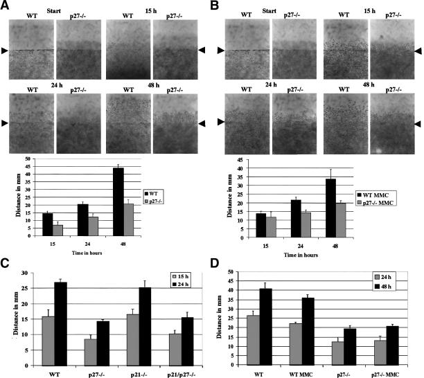
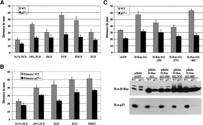
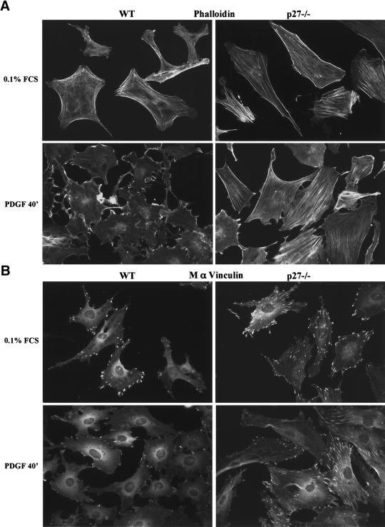
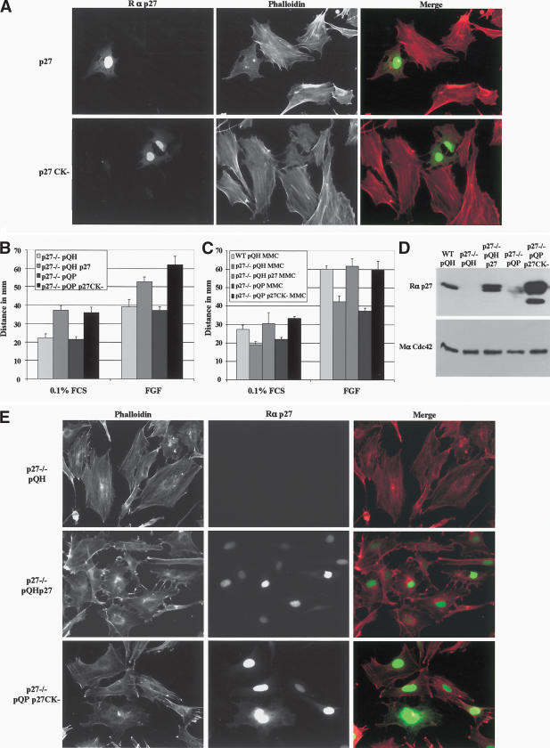
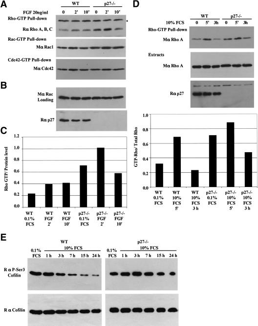
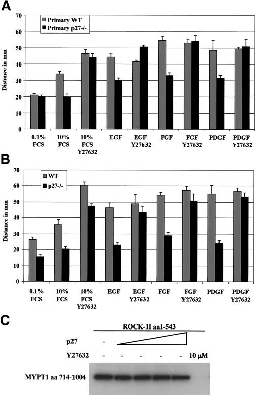
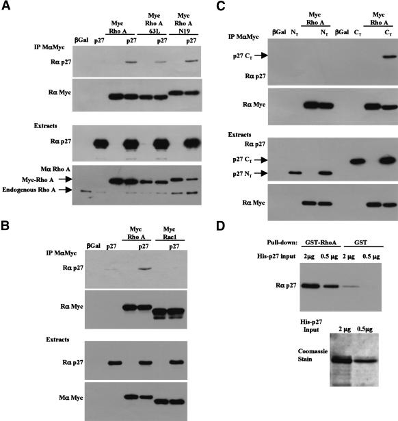
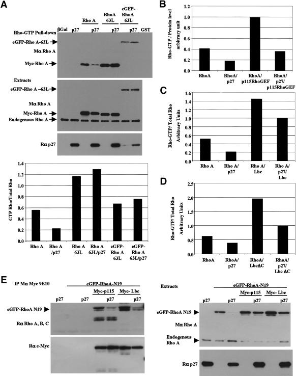
The tumor suppressor p27(Kip1) is an inhibitor of cyclin/cyclin-dependent kinase (CDK) complexes and plays a crucial role in cell cycle regulation. However, p27(Kip1) also has cell cycle-independent functions. Indeed, we find that p27(Kip1) regulates cell migration, as p27(Kip1)-null fibroblasts exhibit a dramatic decrease in motility compared with wild-type cells. The regulation of motility by p27(Kip1) is independent of its cell-cycle regulatory functions, as re-expression of both wild-type p27(Kip1) and a mutant p27(Kip1) (p27CK(-)) that cannot bind to cyclins and CDKs rescues migration of p27(-/-) cells. p27(-/-) cells have increased numbers of actin stress fibers and focal adhesions. This is reminiscent of cells in which the Rho pathway is activated. Indeed, active RhoA levels were increased in cells lacking p27(Kip1). Moreover, inhibition of ROCK, a downstream effector of Rho, was able to rescue the migration defect of p27(-/-) cells in response to growth factors. Finally, we found that p27(Kip1) binds to RhoA, thereby inhibiting RhoA activation by interfering with the interaction between RhoA and its activators, the guanine-nucleotide exchange factors (GEFs). Together, the data suggest a novel role for p27(Kip1) in regulating cell migration via modulation of the Rho pathway.


Figures

Figure 1.
Figure 1.
p27-null MEFs have a migration deficit compared with wild-type MEFs. (A) Migration of immortalized wild-type (WT) and p27–/– MEFs following wounding of a confluent cell monolayer. Migration distances were measured as described in Materials and Methods. (B) Asin A except that cells were pretreated for 3 h with 10μg/mL MMC, to block cell proliferation. (C) Migration of primary wild-type (WT), p27–/–, p21–/–, and p21/p27–/– MEFs. (D) Migration of primary wild-type (WT) and p27–/– MEFs that were treated, or not, with MMC.
Figure 2.
Figure 2.
p27–/– MEFs are refractory to induction of migration by growth factors or activated Ras. (A,B) Migration of immortalized (A) and primary (B) wild-type (WT) and p27–/– MEFs in the presence of growth factors (HGF, FGF2, PDGF-BB, or EGF) at a final concentration of 25 ng/mL. Cells were allowed to migrate for 48 h. (C) Wild-type (WT) and p27–/– were infected with retroviruses expressing the indicated forms of activated Ras. Cells were allowed to migrate for 48 h, and their migration was measured as in Figure 1. (Lower panel) The respective H-Ras and p27Kip1 protein levels of the cells used for the migration assay.
Figure 3
Figure 3
Cells lacking p27Kip1 have increased numbers of actin stress fibers and focal adhesions. Primary MEFs were seeded on glass coverslips, allowed to grow for 16 h, then starved for 48 h in 0.1% FCS, or starved and stimulated for 40 min with 25 ng/mL PDGF-BB (PDGF). (A) Actin was visualized with phalloidin–rhodamine (1/500). (B) Mouse anti-vinculin (1/1000).
Figure 4.
Figure 4.
Re-expression of p27Kip1 or a mutant form that cannot bind to cyclin/CDK (p27CK) rescues the migration and decreases the number of actin stress fibers in p27-null MEFs. (A) Transient transfection of p27–/– MEFs with p27Kip1 (upper row) and p27CK (lower row). Cells were plated on glass coverslips, incubated overnight prior to transfection, and fixed 36 h posttransfection. Cells were stained for p27Kip1 (green) and phalloidin (red). (BE) Immortalized wild-type (WT) and p27–/– MEFs were infected with retroviruses encoding wild-type p27 (pQHp27), or p27CK (pQPp27CK), or the corresponding empty vectors. (B) Migration assays in the presence or absence of 25 ng/mL FGF2. (C) Migration assay with cells pretreated for 3 h with 10 μg/mL MMC. (D) Immunoblot to indicate p27Kip1 levels following retroviral infection in the cells used for the migration assays. The membrane was stripped and reprobed with a monoclonal antibody to Cdc42 to indicate protein loading. Fifty micrograms of protein was loaded per lane. (E) Retrovirally infected p27–/– MEFs were plated on coverslips for 24 h in 10% FCS DMEM, fixed, and stained for p27Kip1 (green) and phalloidin (red).
Figure 5.
Figure 5.
Increased Rho–GTP levels in p27–/– MEFs. (A) Cells were stimulated with FGF2 (25 ng/mL) for the indicated time, and Rho–GTP levels measured using pull-down assays (see Materials and Methods for details). GST-C21 (Rho–GTP) pull-downs were probed with a rabbit anti-RhoA, anti-RhoB, anti-RhoC antibody. GST–PAK1-CD (Rac-GTP and Cdc42–GTP) pull-downs were probed with a mouse anti-Rac1. The membrane was stripped and reprobed with a mouse anti-Cdc42 antibody. The asterisk refers to a nonspecific band in the top panel. (B) A fraction (1/30) of the extracts was used to determine the amount of protein (probed for Rac), and the membrane was reprobed for p27Kip1 after stripping. (C) Quantification of Rho–GTP normalized to the loading control (Rac). (D) Rho–GTP pull-downs with cells starved for 48 h and stimulated with DMEM containing 10% FCS. The graph shows the amount of Rho–GTP in the pull-downs normalized to the total amount of Rho present in the loading control. Similar results were obtained in four independent experiments (E) Increased phosphorylation of cofilin on Ser 3 in p27–/– MEFs. Immortal wild-type (WT) and p27–/– MEFs were starved for 48 h and stimulated for the indicated time with 10% FCS. Ten micrograms of protein was loaded per well. Membranes were probed with a polyclonal antibody to phospho-Ser 3-cofilin, stripped, and reprobed with a polyclonal antibody to cofilin.
Figure 6.
Figure 6.
The Rho-kinase inhibitor Y27632 rescues the migration defect of p27-null MEFs. (A) Migration of primary wild-type (WT) and p27–/– MEFs pretreated for 3 h with 10 μg/mL MMC, and then stimulated with growth factors (FGF2, PDGF-BB, and EGF) at 25 ng/mL in the presence or absence of the Rho-kinase inhibitor Y27632 (10 μM). Cells were allowed to migrate for 48 h. (B) Migration of immortalized wild-type (WT) and p27–/– MEFs, performed as in A except that the cells were not treated with MMC. (C) p27Kip1 does not inhibit ROCK2 in an in vitro kinase assay. Kinase activity of ROCK2 (5.72 nM) was measured in the presence of increasing amounts of recombinant p27Kip1 (2.16 μM, 4.32 μM, 10.81 μM, 21.63 μM, respectively) or the ROCK inhibitor Y27632 (10 μM), using MYPT1 as a substrate.
Figure 7.
Figure 7.
The C-terminal half of p27Kip1 interacts with RhoA in HEK 293T cells. (A) HEK 293T cells were transfected with full-length p27Kip1 and/or Myc-tagged RhoA constructs. RhoA was immunoprecipitated using a monoclonal Myc antibody (9E10). The immunoprecipitates were probed with a p27Kip1 antibody (rabbit, C19; top panel), and the membrane was stripped and reprobed for Myc (rabbit, A14) to indicate the amount of myc-RhoA immunoprecipitated. The amounts of p27Kip1 and RhoA in the cell extracts are shown in the lower panels. (B) As in A except that cells were transfected with p27Kip1 and/or Myc-tagged RhoA or Myc-tagged Rac1 to test the specificity of the interaction of p27 with RhoA. (C) As in A except the N-terminal half (amino acids 1–85, NT) or the C-terminal half (amino acids 86–198, CT) of p27Kip1 were cotransfected with Myc-RhoA. Only the C-terminal half of p27Kip1 was detected with a mix of N- and C-terminal p27Kip1 antibodies (rabbit, C19 and N20) in the Myc immunoprecipitates. A fraction of the lysates (1/12th of the amount used for immunoprecipitations) was probed for p27Kip1 and Myc to indicate the levels of the transfected proteins (lower panel). (D) In vitro binding assay. The indicated amounts of recombinant His-tagged p27Kip1 were incubated with an excess of GST–RhoA or GST beads. The amount of His-p27Kip1 recovered in the pull-downs was detected by immunoblot using a p27Kip1 antibody. (Lower panel) The amounts of His-p27Kip1 used in the pull-downs, stained with Coomassie blue.
Figure 8.
Figure 8.
p27Kip1 inhibits RhoA activation by interfering with the binding of RhoA to its GEFs. (A) p27Kip1 does not affect the Rho/effector interaction. HEK 293T cells were cotransfected with p27Kip1 and the indicated RhoA expression vectors. (Top panel) The amount of Rho–GTP was determined in a pull-down assay, as described (see Fig. 5; Materials and Methods). (Bottom two panels) The amount of RhoA and p27Kip1 in the cellular extracts was determined by immunoblot. The graph represents the amount of GTP–RhoA normalized to the total amount of transfected RhoA protein (GTP–Rho/Total Rho). Similar results were obtained in three independent experiments. (BD) p27Kip1 interferes with the activation of RhoA by its GEFs. As in A, except that cells were cotransfected with Myc-RhoA and/or p27Kip1, and/or Myc-p115RhoGEF (B), Myc-Lbc (C), and Myc-LbcΔC (D). The graphs indicate the amount of GTP–RhoA normalized to the total amount of transfected RhoA protein. For each GEF, similar results were obtained in at least two independent experiments. (E) p27Kip1 inhibits RhoA binding to its GEFs. HEK 293T cells were cotransfected with the indicated expression vectors. (Left panel, top) The Myc-tagged GEFs were immunoprecipitated using a Myc antibody (mouse, 9E10), and the amount of eGFP–RhoA-N19 coprecipitated with the GEFs was determined by immunoblot. Note that the lower band in the Myc-p115 lanes are nonspecific. The membrane was reprobed with a Myc antibody (rabbit, A14) to indicate the amount of GEF immunoprecipitated. The expression levels of eGFP–RhoA-N19 and p27Kip1 are indicated on the right panel (note that expression of eGFP–RhoA-N19 was very low in the control lane 3). Similar results were obtained in four independent experiments.

References

    1. Amano, A., Chihara, K., Kimura, K., Fukata, Y., Nakamura, N., Matsuura, Y., and Kaibuchi, K. 1997. Formation of actin stress fibers and focal adhesions enhanced by rho-kinase. Science 275: 1308–1311. - PubMed
    1. Anayama, T., Furihata, M., Ishikawa, T., Ohtsuki, Y., and Ogoshi, S. 1998. Positive correlation between p27Kip1 expression and progression in human esophageal squamous cell carcinoma. Int. J. Cancer 79: 439–443. - PubMed
    1. Arthur, W.T. and Burridge, K. 2001. RhoA inactivation by p190RhoGAP regulates cell spreading and migration by promoting membrane protrusion and polarity. Mol. Biol. Cell 12: 2711–2720. - PMC - PubMed
    1. Arthur, W.T., Petch, L.A., and Burridge, K. 2000. Integrin engagement suppresses RhoA activity via a c-Src-dependent mechanism. Curr. Biol. 10: 719–722. - PubMed
    1. Bar-Sagi, D. and Hall, A. 2000. Ras and Rho GTPases: A family reunion. Cell 103: 227–238. - PubMed

Publication types

MeSH terms