. 2004 Apr 15;18(8):877-88.
doi: 10.1101/gad.1184604. Epub 2004 Apr 12.

Nucleosomal histone kinase-1 phosphorylates H2A Thr 119 during mitosis in the early Drosophila embryo

Affiliations

Nucleosomal histone kinase-1 phosphorylates H2A Thr 119 during mitosis in the early Drosophila embryo

Hitoshi Aihara et al. Genes Dev. .

Abstract

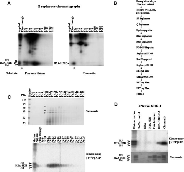
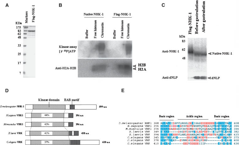
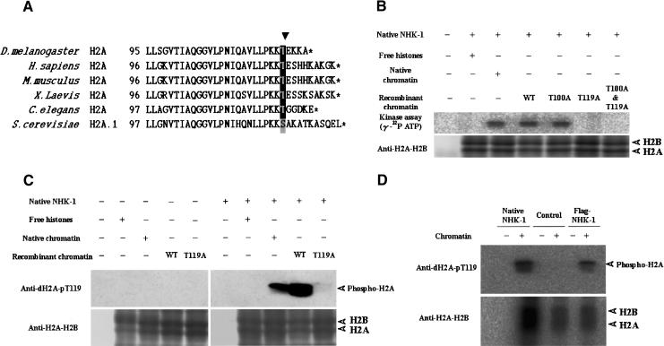
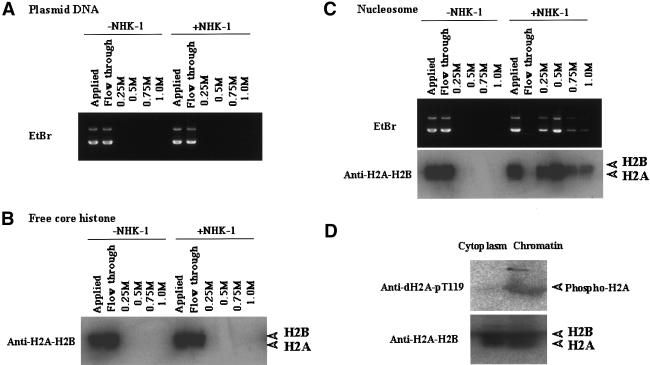
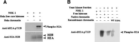
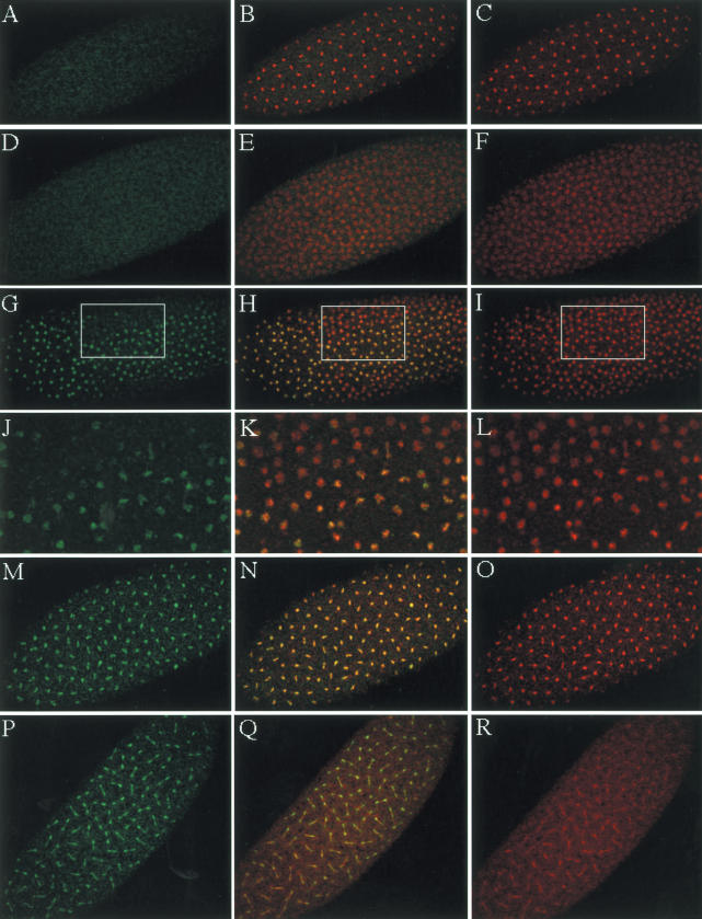
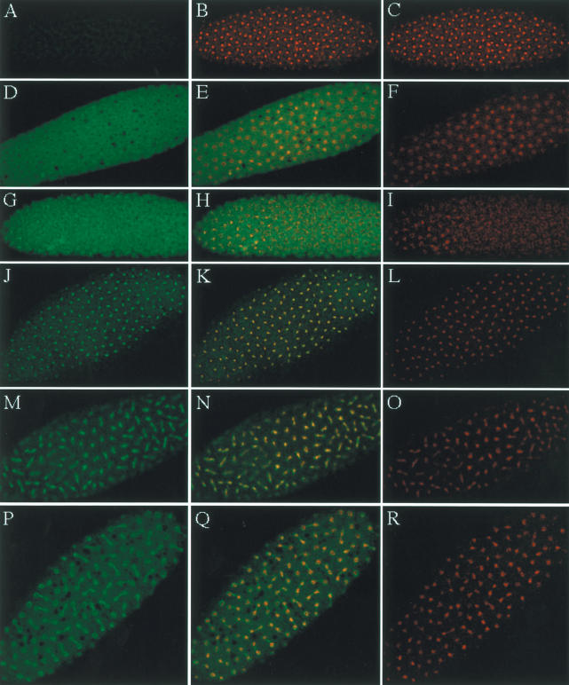
Posttranslational histone modifications are important for the regulation of many biological phenomena. Here, we show the purification and characterization of nucleosomal histone kinase-1 (NHK-1). NHK-1 has a high affinity for chromatin and phosphorylates a novel site, Thr 119, at the C terminus of H2A. Notably, NHK-1 specifically phosphorylates nucleosomal H2A, but not free H2A in solution. In Drosophila embryos, phosphorylated H2A Thr 119 is found in chromatin, but not in the soluble core histone pool. Immunostaining of NHK-1 revealed that it goes to chromatin during mitosis and is excluded from chromatin during S phase. Consistent with the shuttling of NHK-1 between chromatin and cytoplasm, H2A Thr 119 is phosphorylated during mitosis but not in S phase. These studies reveal that NHK-1-catalyzed phosphorylation of a conserved serine/threonine residue in H2A is a new component of the histone code that might be related to cell cycle progression.


Figures

Figure 1.
Figure 1.
Identification and purification of nucleosomal histone kinase-1 (NHK-1). (A) Kinase assay from Q Sepharose chromatography (see B). Fractions were incubated with free core histones or chromatin as substrates in the presence of [γ-32P]ATP. These reaction mixtures were run on 15% SDS-PAGE and analyzed by autoradiography. Asterisks indicate initial identification of nucleosomal histone kinase activity. (B) Scheme for purification of NHK-1. (C, bottom panel) The kinase assay using both chromatin as substrate and fractions from the final HiTrap blue column chromatography (see B). (Top panel) These same fractions were precipitated with TCA, run on 10% SDS-PAGE, and stained with Coomassie Brilliant Blue. The p70, p52, and p42 bands indicated by asterisks were subjected to peptide sequencing. (D) Kinase assay used highly purified native NHK-1 in the presence of DNA, H2A–H2B dimer, all four core histones, H2A–H2B/DNA, or chromatin as substrates. All four core histones were phosphorylated by nonspecific kinase and used as histone markers.
Figure 2.
Figure 2.
Purified recombinant NHK-1 phosphorylates nucleosomal H2A–H2B. (A) Expression of recombinant Flag-tagged NHK-1 by E. coli. (B) Recombinant NHK-1 phosphorylates H2A–H2B in chromatin form. Kinase assay used native NHK-1 and Flag-tagged recombinant NHK-1 in the presence of free core histones or chromatin and [γ-32P]ATP. For quantification of nucleosomes, anti-dH2A-H2B was used. (C) Equivalent amounts of the protein extracts (20 μg) derived from Drosophila embryos before and after gastrulation were subjected to SDS-PAGE and Western blot analysis together with Flag-NHK-1. (Upper panel) The presence of NHK-1 detected with anti-NHK-1. (Lower panel) The presence of dNLP, which was proven to be expressed throughout development, to confirm the recovery of total protein (Ito et al. 1996a). (D) Schematic diagram of NHK-1 protein and its homologs; Homo sapiens VRK1 (Vaccinia related kinase 1), Mus musculus VRK1, Xenopus laevis VRK, and Caenorhabditis elegans VRK. The percent identity between each kinase and dNHK-1 over the kinase domain is shown in the boxes. (E) Alignment of basic-acidic-basic amino acid motif (BAB motif) in NHK-1 and counterparts of other homologs. Acidic and basic amino acids are shaded red or blue, respectively.
Figure 3.
Figure 3.
NHK-1 specifically phosphorylates Thr 119 of the histone H2A C terminus in vitro. (A) Sequence alignment of H2A C termini from Drosophila melanogaster, Homo sapiens, Mus musculus, Xenopus laevis, Caenorhabditis elegans, and Saccharomyces cerevisiae. The amino acid position in each protein is shown on the left. Stop codons are indicated by asterisks. The arrowhead indicates evolutionally conserved serine/threonine. (B) Drosophila native NHK-1 phosphorylates H2A Thr 119 specifically. A kinase assay was performed in the presence of [γ-32P]ATP using chromatin reconstituted with native core histones purified from Drosophila or recombinant core histones expressed by E. coli. (C) Antibodies raised against the phospho-Thr 119 H2A peptide specifically recognize phosphorylation of Drosophila Thr 119 of H2A. Kinase assay was done in the presence of core histones or equivalent amount of chromatin. (D) Recombinant NHK-1 also phosphorylates Thr 119 of H2A. Chromatin was subjected to kinase assay using recombinant NHK-1 and native NHK-1. As control, partially purified nonspecific histone kinase, which phosphorylates only free core histones, was used. Phosphorylation was detected by Western blotting with anti-dH2A-pT119. T100A or T119A indicates that H2A Thr 100 or Thr 119 is substituted with alanine, respectively. (B,C) WT indicates chromatin reconstituted with wild-type H2A. (BD) Anti-dH2A-H2B was used for quantification of nucleosomes.
Figure 4.
Figure 4.
NHK-1 has higher affinity for chromatin than free histones. Recombinant NHK-1 was immobilized to resin and incubated with DNA (A), free core histones (B), or chromatin (C). Affinity was examined by stepwise salt elution with 0.25, 0.50, 0.75, and 1.0 M KCl. Each sample was analyzed by Western blotting with anti-H2A–H2B or agarose gel electrophoresis followed by EtBr staining. (D) Thr 119 of histone H2A is phosphorylated in chromatin form but not in the free histone pool in vivo. The cytoplasmic fraction and chromatin fraction from 0–1-h Drosophila embryos were subjected to Western blotting with anti-dH2A-pT119 and anti-dH2A–H2B.
Figure 5.
Figure 5.
NHK-1 activity is conserved among species. (A) Recombinant dNHK-1 can phosphorylate human chromatin. The reaction used recombinant human NHK-1 and chromatin reconstituted with HeLa core histones or HeLa core histones only. The reaction mixture was subjected to Western blotting after incubation with cold ATP. (B) Yeast NHK phosphorylates Drosophila Thr 119 of H2A only in chromatin form. Partially purified yeast NHK with SP Sepharose chromatography and native dNHK-1 were subjected to kinase assay with anti-dH2A-pT119.
Figure 6.
Figure 6.
Histone H2A Thr 119 is phosphorylated during mitosis. (A,D,G,J,M,P) For immunostaining, 0–1-h Drosophila embryos were used. Drosophila embryos were immunostained with anti-dH2A-pT119. (C,F,I,L,O,R) DNA was visualized with Actinomycin D. (B,E,H,K,N,Q) Immunostaining with rabbit anti-dH2A-pT119 and DNA staining were merged, and overlapping is visualized by yellow. Immunostaining of S phase (DF), prophase (GL), metaphase (MO), and anaphase and telophase (PR) are shown. (AC) Phosphopeptide was used to block anti-dH2A-pT119 as control. There was no significant nuclear staining when antibody is blocked with phosphopeptide as shown in A. Enlargements of the boxes in G, H, and I are shown in J, K, and L, respectively.
Figure 7.
Figure 7.
The localization of NHK-1 in the syncytial Drosophila embryo is changing depending on cell cycle. For immunostaining, 0–1-h Drosophila embryos were used. Drosophila embryos were immunostained with preimmune serum (A) and rabbit anti-NHK-1 antibody (D,G,J,M,P). (C,F,I,L,O,R) DNA was visualized with Actinomycin D. (B,E,H,K,N,ITLQ) Immunostaining with preimmune serum or anti-NHK-1 and DNA staining were merged, and overlapping is visualized by yellow. Immunostaining of S phase (DF), prophase (GI), metaphase (JO), and anaphase and telophase (PR) is shown.

Similar articles

Cited by

References

    1. Anest, V., Hanson, J.L., Cogswell, P.C., Steinbrecher, K.A., Strahl, B.D., and Baldwin, A.S. 2003. A nucleosomal function for IκB kinase-α in NF-κB-dependent gene expression. Nature 423: 659–663. - PubMed
    1. Ballal, N.R., Kang, Y.J., Olson, M.O., and Busch, H. 1975. Changes in nucleolar proteins and their phosphorylation patterns during liver regeneration. J. Biol. Chem. 250: 5921–5925. - PubMed
    1. Bannister, A.J., Zegerman, P., Partridge, J.F., Miska, E.A., Thomas, J.O., Allshire, R.C., and Kouzarides, T. 2001. Selective recognition of methylated lysine 9 on histone H3 by the HP1 chromo domain. Nature 410: 120–124. - PubMed
    1. Carrozza, M.J., Utley, R.T., Workman, J.L., and Cote, J. 2003. The diverse functions of histone acetyltransferase complexes. Trends Genet. 19: 321–329. - PubMed
    1. Cheung, P., Tanner, K.G., Cheung, W.L., Sassone-Corsi, P., Denu, J.M., and Allis, C.D. 2000. Synergistic coupling of histone H3 phosphorylation and acetylation in response to epidermal growth factor stimulation. Mol. Cell 5: 905–915. - PubMed