. 2004 Apr 19;199(8):1089-99.
doi: 10.1084/jem.20040051. Epub 2004 Apr 12.

TOX provides a link between calcineurin activation and CD8 lineage commitment

Affiliations

TOX provides a link between calcineurin activation and CD8 lineage commitment

Parinaz Aliahmad et al. J Exp Med. .

Abstract

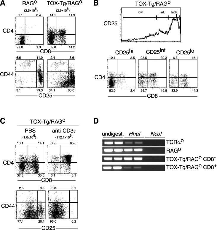
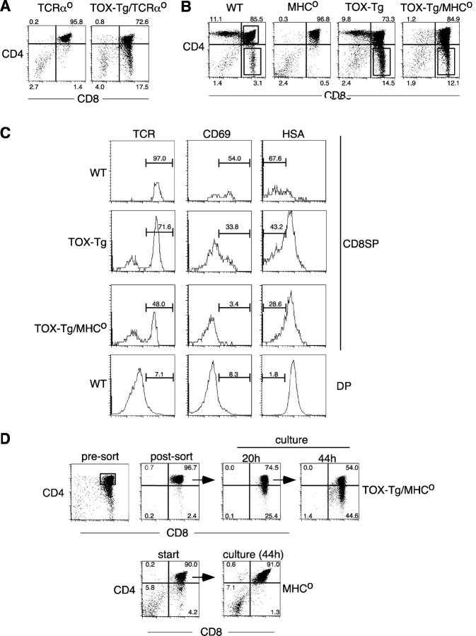
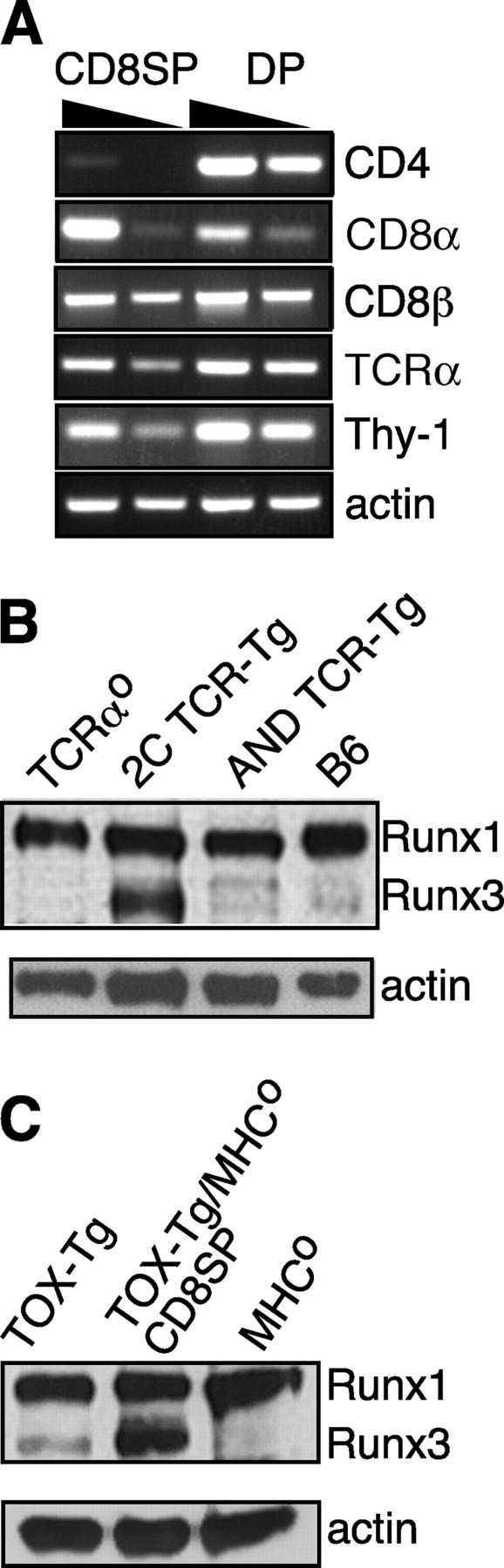
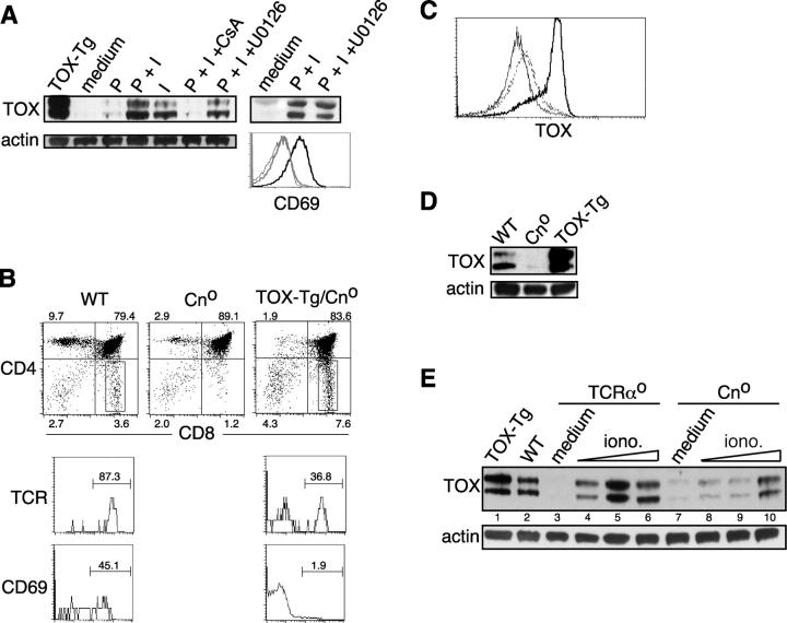
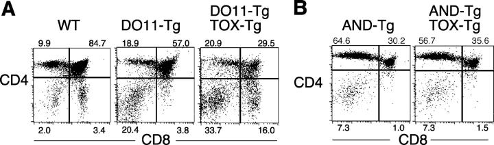
T cell development is dependent on the integration of multiple signaling pathways, although few links between signaling cascades and downstream nuclear factors that play a role in thymocyte differentiation have been identified. We show here that expression of the HMG box protein TOX is sufficient to induce changes in coreceptor gene expression associated with beta-selection, including CD8 gene demethylation. TOX expression is also sufficient to initiate positive selection to the CD8 lineage in the absence of MHC-TCR interactions. TOX-mediated positive selection is associated with up-regulation of Runx3, implicating CD4 silencing in the process. Interestingly, a strong T cell receptor-mediated signal can modify this cell fate. We further demonstrate that up-regulation of TOX in double positive thymocytes is calcineurin dependent, linking this critical signaling pathway to nuclear changes during positive selection.


Figures

Figure 1.
Figure 1.
TOX induces development of coreceptor positive thymocytes in RAGo mice. (A) TOX-Tg/RAGo thymocytes were stained for expression of CD4, CD8α, CD44, and CD25 and analyzed by flow cytometry. Percentages of total thymocytes in each quadrant and total thymic cellularity are indicated. (B) CD4 and CD8α expression (dot plots) on CD25 gated thymocytes (histogram) in a TOX-Tg/RAGo mouse. (C) TOX-Tg/RAGo mice were injected i.p. with PBS or anti-CD3ɛ antibody, and the thymocytes were analyzed 5 d later for expression of CD4 and CD8α or CD44 and CD25 as in A. (D) Methylation status of the CD8 locus in thymocytes from mice with the indicated genotype as analyzed by restriction digest and PCR. CD8α+ and CD8α2 thymocyte cell subpopulations were purified by cell sorting.
Figure 2.
Figure 2.
TOX induces development of CD8SP thymocytes in the absence of TCR–MHC interactions. (A and B) Expression of CD4 and CD8α on thymocytes from mice with the indicated genotype. Percentages of total thymocytes in each quadrant are indicated. (C) Expression of TCRβ, CD69, and HSA on CD8SP thymocytes gated as in B from mice with the indicated genotypes. DP thymocytes from wild-type mice were used as a control to set appropriate gates. The percentage of TCRhi, CD69+, and HSAlo cells are shown. (D) Total MHCo thymocytes or sorted DP thymocytes from TOX-Tg/MHCo mice were cultured ex vivo and analyzed for CD4 and CD8α expression by flow cytometry after 20 or 44 h. Percentages of total thymocytes in each quadrant are shown. Viable yield of TOX-Tg/MHCo thymocytes was 73% after 20 h of culture.
Figure 3.
Figure 3.
CD4 gene silencing in TOX-Tg mice. (A) RT-PCR analysis for expression of the indicated genes in purified CD8SP or DP thymocytes isolated from TOX-Tg/MHCo mice. (B and C) Western blot analysis for expression of Runx proteins in whole cell lysates prepared from total thymocytes or isolated CD8SP cells as indicated. Expression of β-actin was used as a loading control.
Figure 4.
Figure 4.
Expression of TOX is regulated by Cn activation. (A) Expression of TOX in TCRαo thymocytes activated with PMA and ionomycin in the absence or presence of inhibitors of Cn (cyclosporin A, CsA) or MEK (U0126). Also shown is CD69 expression on cells cultured in medium (thin line), with PMA and ionomycin (thick black line), or with PMA/ionomycin and U0126 (thick gray line). Expression of TOX in thymocytes derived from a TOX-Tg mouse is shown as a control. (B) Expression of CD4 and CD8α on thymocytes derived from various animals as indicated (dot plots). Percentage of total thymocytes in each quadrant is shown. Expression of TCRβ and CD69 was also analyzed on gated CD8SP thymocytes, and the percentage of TCRhi or CD69+ cells is indicated. Expression of TOX was determined by intracellular staining and flow cytometry (C) in WT (dashed line), Cno (thin line), and TOX-Tg (thick line) thymocytes or by Western blot of whole cell lysates (D). (E) Thymocytes from TCRαo and Cno mice were cultured in medium (lanes 3 and 7) or 0.2 ng/ml of PMA and 0.05 (lanes 4 and 8), 0.1 (lanes 5 and 9), or 0.2 (lanes 6 and 10) μg/ml of ionomycin. TOX-Tg thymocytes were analyzed as a control (lane 1).
Figure 5.
Figure 5.
TCR signaling in vivo competes with TOX activity. (A and B) Expression of CD4 and CD8α on thymocytes from indicated mice was analyzed by flow cytometry. Percentage of total thymocytes in each quadrant is shown.

Similar articles

Cited by

References

    1. Starr, T.K., S.C. Jameson, and K.A. Hogquist. 2003. Positive and negative selection of T cells. Annu. Rev. Immunol. 21:139–176. - PubMed
    1. van Oers, N.S. 1999. T cell receptor-mediated signs and signals governing T cell development. Semin. Immunol. 11:227–237. - PubMed
    1. Kioussis, D., and W. Ellmeier. 2002. Chromatin and CD4, CD8A and CD8B gene expression during thymic differentiation. Nat. Rev. Immunol. 2:909–919. - PubMed
    1. Harker, N., T. Naito, M. Cortes, A. Hostert, S. Hirschberg, M. Tolaini, K. Roderick, K. Georgopoulos, and D. Kioussis. 2002. The CD8alpha gene locus is regulated by the Ikaros family of proteins. Mol. Cell. 10:1403–1415. - PubMed
    1. Taniuchi, I., M. Osato, T. Egawa, M.J. Sunshine, S.C. Bae, T. Komori, Y. Ito, and D.R. Littman. 2002. Differential requirements for Runx proteins in CD4 repression and epigenetic silencing during T lymphocyte development. Cell. 111:621–633. - PubMed

Publication types

MeSH terms