. 2004 Apr 19;199(8):1041-52.
doi: 10.1084/jem.20031080. Epub 2004 Apr 12.

c-FLIP mediates resistance of Hodgkin/Reed-Sternberg cells to death receptor-induced apoptosis

Affiliations

c-FLIP mediates resistance of Hodgkin/Reed-Sternberg cells to death receptor-induced apoptosis

Stephan Mathas et al. J Exp Med. .

Abstract

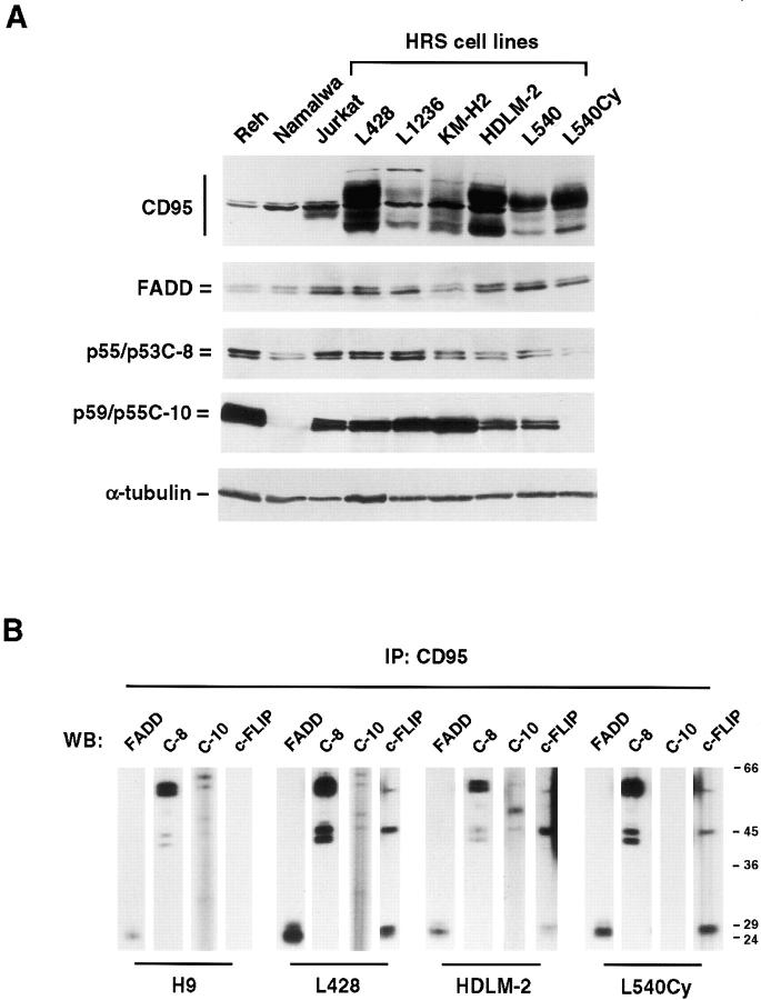
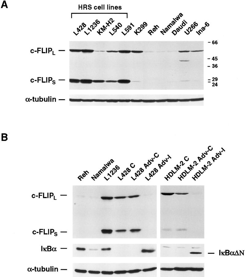
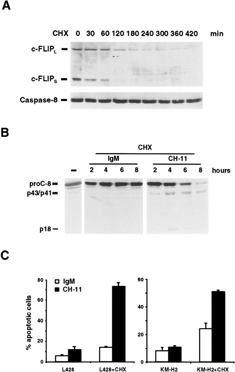
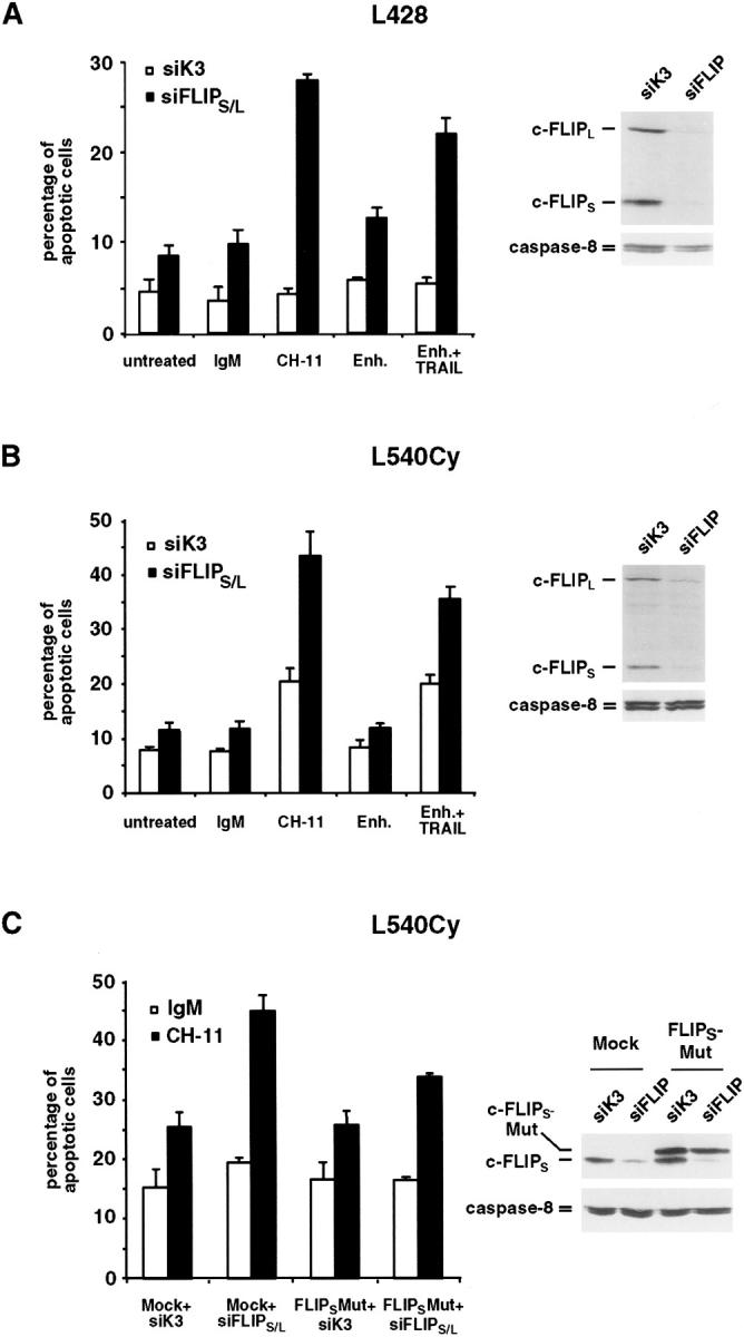
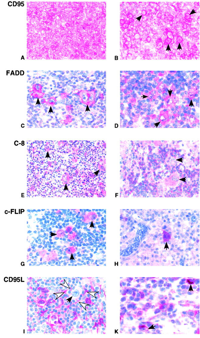
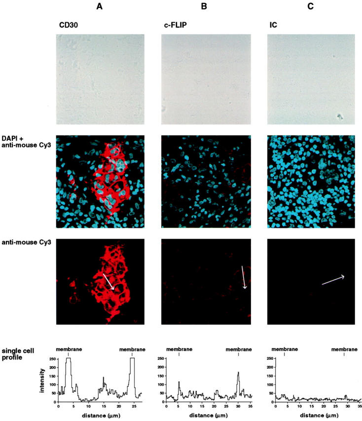
Resistance to death receptor-mediated apoptosis is supposed to be important for the deregulated growth of B cell lymphoma. Hodgkin/Reed-Sternberg (HRS) cells, the malignant cells of classical Hodgkin's lymphoma (cHL), resist CD95-induced apoptosis. Therefore, we analyzed death receptor signaling, in particular the CD95 pathway, in these cells. High level CD95 expression allowed a rapid formation of the death-inducing signaling complex (DISC) containing Fas-associated death domain-containing protein (FADD), caspase-8, caspase-10, and most importantly, cellular FADD-like interleukin 1beta-converting enzyme-inhibitory protein (c-FLIP). The immunohistochemical analysis of the DISC members revealed a strong expression of CD95 and c-FLIP overexpression in 55 out of 59 cases of cHL. FADD overexpression was detectable in several cases. Triggering of the CD95 pathway in HRS cells is indicated by the presence of CD95L in cells surrounding them as well as confocal microscopy showing c-FLIP predominantly localized at the cell membrane. Elevated c-FLIP expression in HRS cells depends on nuclear factor (NF)-kappaB. Despite expression of other NF-kappaB-dependent antiapoptotic proteins, the selective down-regulation of c-FLIP by small interfering RNA oligoribonucleotides was sufficient to sensitize HRS cells to CD95 and tumor necrosis factor-related apoptosis-inducing ligand-induced apoptosis. Therefore, c-FLIP is a key regulator of death receptor resistance in HRS cells.


Figures

Figure 1.
Figure 1.
DISC analysis in HRS cell lines. (A) Whole cell lysates of different nonHodgkin and Hodgkin cell lines were analyzed for expression of CD95, FADD, C-8 (p55/p53C-8), and C-10 (p59/p55C-10) by Western blot. As control, α-tubulin expression is shown. (B) DISC analysis in H9 cells and HRS cell lines by CD95 immunoprecipitation. The indicated cell lines were incubated for 10 min with the agonistic anti-CD95 mAb APO-1. Thereafter, cells were lysed and immunoprecipitation was performed. The precipitates were washed, subjected to 10% SDS-PAGE, and analyzed by Western blot for coimmunoprecipitation of FADD, C-8, C-10, and c-FLIP, as indicated.
Figure 2.
Figure 2.
c-FLIP expression and regulation in HRS cell lines. (A) Elevated c-FLIP protein expression in HRS compared with nonHodgkin cell lines. Whole cell lysates of the indicated cell lines were analyzed by Western blot for expression of c-FLIPL and c-FLIPS (mAb Dave-2). As control, the Western blot was reprobed with an α-tubulin antibody. (B) c-FLIP expression in HRS cell lines depends on NF-κB. Whole cell extracts of Reh, Namalwa, L1236, uninfected L428 (L428 C), and HDLM-2 (HDLM-2 C) cells, and L428 or HDLM-2 cells infected for 24 h with a control adenovirus (L428 Adv-C, HDLM-2 Adv-C) or an adenovirus encoding for the NF-κB superrepressor IκBαΔN (L428 Adv-I, HDLM-2 Adv-I) were analyzed by Western blot analysis for expression of c-FLIP, IκBα, IκBαΔN, and as control, α-tubulin. Note that L428 cells lack endogenous IκBα expression.
Figure 3.
Figure 3.
Treatment with CHX down-regulates c-FLIP and sensitizes HRS cells to CD95-induced apoptosis. (A) L428 cells were treated with CHX. At the indicated times, whole cell lysates were prepared and analyzed for expression of c-FLIPL and c-FLIPS and C-8 by Western blot. (B) Treatment with CHX allows CD95-induced activation of C-8. L428 cells were left untreated (−) or incubated with CHX. After 2 h, the agonistic anti-CD95 antibody CH-11 or an isotype control (IgM) were added to the CHX-treated cells. At the indicated times, whole cell lysates were prepared and analyzed for expression of C-8 by Western blot. Positions of procaspase-8 (proC-8) and cleavage products (p43/p41, p18) are indicated. (C) Treatment of HRS cell lines with CH-11 induces apoptosis in the presence of CHX. L428 and KM-H2 cells were treated for 14 h with CHX together with the agonistic anti-CD95 antibody CH-11 or an IgM control. The percentage of apoptotic cells was determined by acridine orange staining. Error bars denote SDs.
Figure 4.
Figure 4.
Specific down-regulation of c-FLIP sensitizes HRS cell lines to CD95- and TRAIL-induced apoptosis. (A) L428 cells were transfected with control (siK3) or siFLIPS/L along with pEGFP-N3. 24 h after transfection, GFP+ cells were FACS® sorted, analyzed for expression of c-FLIP by Western blot (right; as control, analysis of C-8 is shown), and left untreated or were incubated for 14 h with an IgM control (IgM) or the agonistic anti-CD95 antibody CH-11, cross-linking antibody for TRAIL alone (enhancer, Enh.), or with enhancer and recombinant human soluble TRAIL (Enh. + TRAIL). Thereafter, the percentage of apoptotic cells was determined by acridine orange staining (left). Error bars denote SDs. (B) L540Cy cells were transfected and analyzed for c-FLIP and C-8 expression (right) as described in A. 24 h after transfection, cells were left untreated or treated as described in A. Thereafter, the percentage of apoptotic cells was determined by annexin V–FITC and propidium iodide staining and subsequent FACS® analysis (left). The combined results of four independent experiments are shown. Error bars denote SDs. (C) L540Cy cells were transfected with control (siK3) or siFLIPS/L along with mock plasmid (Mock) or a mutated c-FLIPS cDNA (FLIPS-Mut), which contains seven silent mutations and cannot be recognized by siFLIP. 24 h after transfection, cells were analyzed for endogenous (c-FLIPS) or ectopically expressed c-FLIP (c-FLIPS-Mut) and C-8 by Western blot (right), and further processed as described in B. The combined results of three independent experiments are shown (left). Error bars denote SDs.
Figure 5.
Figure 5.
Expression patterns of CD95, FADD, C-8, c-FLIP, and CD95L in HRS cells of cHL. Immunohistochemistry of representative biopsy specimen. (A and B) Strong CD95 expression in HRS cells (exemplarily marked by arrows). (C) Strong FADD expression in HRS (arrows), but not in surrounding cells. (D) Moderate FADD expression in HRS cells (arrows). (E and F) C-8 expression of two representative cases. (E) HRS cells (arrows) stain at least as strong as surrounding cells for C-8. (F) HRS cells stain in a less intense fashion than surrounding cells. (G and H) Strong c-FLIP expression in HRS cells (arrows), but not surrounding cells. (I and K) CD95L expression in cHL. (I) In the majority of cases, HRS cells (solid arrow) do not stain for CD95L. In contrast, surrounding cells (open arrows) stain for CD95L. (K) In one case, strong expression of CD95L was detectable in HRS cells (arrows).
Figure 6.
Figure 6.
Confocal microscopy of c-FLIP and CD30 in a representative biopsy specimen. Biopsy specimen were stained with CD30 (A), c-FLIP (B), or isotype control (IC; C) mAbs. Detection was performed after staining with an anti–mouse Cy3 conjugate. Nuclei were counterstained with DAPI. For each antibody, the staining intensity for a representative single HRS cell is shown as single cell profile. Localization of the profile is indicated by a white arrow.

References

    1. Locksley, R.M., N. Killeen, and M.J. Lenardo. 2001. The TNF and TNF receptor superfamilies: integrating mammalian biology. Cell. 104:487–501. - PubMed
    1. Peter, M.E., and P.H. Krammer. 2003. The CD95(APO-1/Fas) DISC and beyond. Cell Death Differ. 10:26–35. - PubMed
    1. Smyth, M.J., K. Takeda, Y. Hayakawa, J.J. Peschon, M.R. van den Brink, and H. Yagita. 2003. Nature's TRAIL–on a path to cancer immunotherapy. Immunity. 18:1–6. - PubMed
    1. Irmler, M., M. Thome, M. Hahne, P. Schneider, K. Hofmann, V. Steiner, J.L. Bodmer, M. Schroter, K. Burns, C. Mattmann, et al. 1997. Inhibition of death receptor signals by cellular FLIP. Nature. 388:190–195. - PubMed
    1. Thome, M., and J. Tschopp. 2001. Regulation of lymphocyte proliferation and death by FLIP. Nat. Rev. Immunol. 1:50–58. - PubMed

Publication types

MeSH terms

Substances