. 2004 Apr 19;199(8):1065-75.
doi: 10.1084/jem.20022212. Epub 2004 Apr 12.

Blockade of attachment and fusion receptors inhibits HIV-1 infection of human cervical tissue

Affiliations

Blockade of attachment and fusion receptors inhibits HIV-1 infection of human cervical tissue

Qinxue Hu et al. J Exp Med. .

Abstract

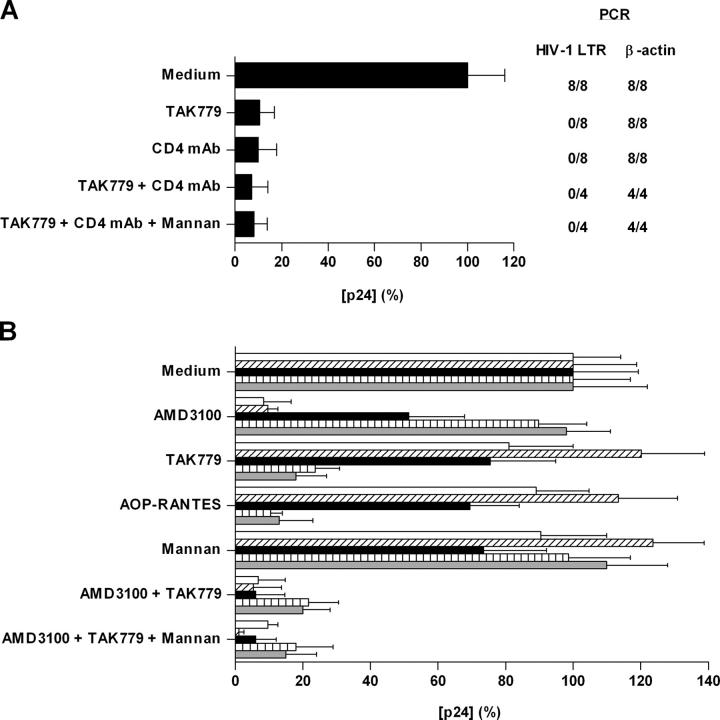
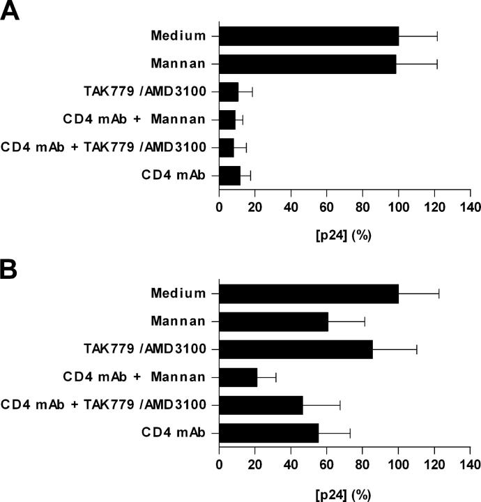
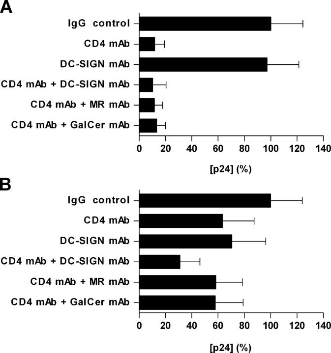
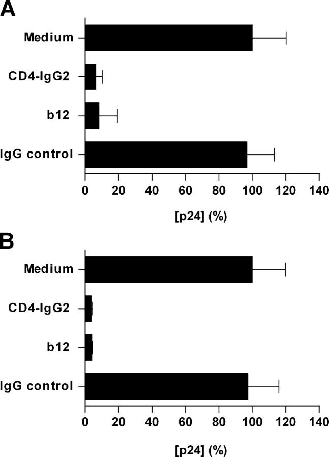
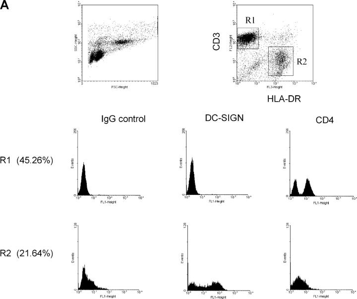
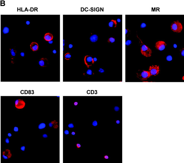
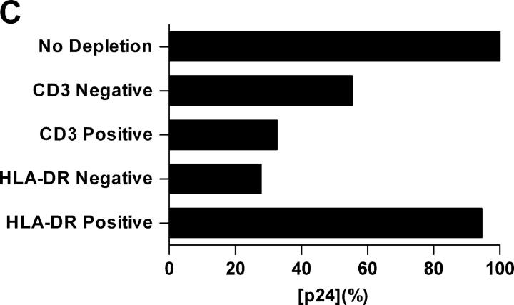
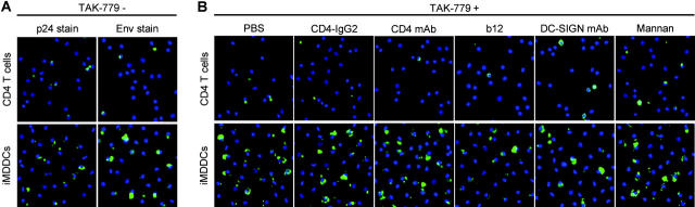
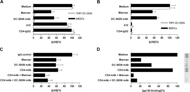
Identification of cellular factors involved in HIV-1 entry and transmission at mucosal surfaces is critical for understanding viral pathogenesis and development of effective prevention strategies. Here we describe the evaluation of HIV-1 entry inhibitors for their ability to prevent infection of, and dissemination from, human cervical tissue ex vivo. Blockade of CD4 alone or CCR5 and CXCR4 together inhibited localized mucosal infection. However, simultaneous blockade of CD4 and mannose-binding C-type lectin receptors including dendritic cell-specific intercellular adhesion molecule-grabbing integrin was required to inhibit HIV-1 uptake and dissemination by migratory cells. In contrast, direct targeting of HIV-1 by neutralizing mAb b12 and CD4-IgG2 (PRO-542) blocked both localized infection and viral dissemination pathways. Flow cytometric analysis and immunostaining of migratory cells revealed two major populations, CD3(+)HLA-DR(-) and CD3(-)HLA-DR(+) cells, with a significant proportion of the latter also expressing dendritic cell-specific intercellular adhesion molecule-grabbing integrin. Bead depletion studies demonstrated that such HLA-DR(+) cells accounted for as much as 90% of HIV-1 dissemination. Additional studies using immature monocyte-derived dendritic cells demonstrated that although mannose-binding C-type lectin receptors and CD4 are the principal receptors for gp120, other mechanisms may account for virus capture. Our identification of the predominant receptors involved in HIV-1 infection and dissemination within human cervical tissue highlight important targets for microbicide development.


Figures

Figure 1.
Figure 1.
Receptor blockade inhibits HIV-1 infection of cervical explant tissue. (A) Unstimulated or (B) PHA-activated cervical explants were incubated with inhibitors for 1 h at 37°C before exposure to (A) HIV-1BaL or (B) NL4–3, 2044, 2076, SL2, and BaL for 2 h at 37°C. After incubation, explants were extensively washed and cultured at 37°C for 14 d. Virus production in the absence of inhibitor was defined as 100% and was (A) 1.92 ng/ml or (B) 1.39 (NL4.3, open bar), 1.04 (2044, hatched bar), 0.94 (2076, black bar), 0.79 (SL2, vertical hatched bar), and 1.70 ng/ml (BaL, gray bar). The data shown are representative of four independent experiments from separate donors yielding similar results, with each p24 determination shown as the mean (± SD) of triplicate determinations. Proviral DNA accumulation (A) at day 14 was determined by quantitative real-time PCR using LTR primers. β-Actin primers were used to control for total amount of DNA.
Figure 2.
Figure 2.
Inhibition of localized mucosal HIV-1 infection and dissemination pathways. Human cervical explants were incubated with inhibitors for 1 h at 37°C before exposure to HIV-1BaL for 2 h at 37°C. After incubation, explants were extensively washed and cultured in the presence of 100 ng/ml of MIP-3β for 48 h. Emigrated wells were collected, washed, and cocultured with PM1 cells. The explants were cultured in separate wells. The data shown represent mean p24 antigen release (± SD) from both (A) cultured explants and (B) cocultured migratory cells and were derived from three separate donors with each determination performed in triplicates. Virus production in the absence of inhibitor was defined as 100% and was 0.99 and 1.26 ng/ml for the cervical explants and migratory cells, respectively.
Figure 3.
Figure 3.
Multiple receptors are involved in HIV-1 uptake by migratory cells. Experiments were performed as described in the legend for Fig. 2. The data shown represent mean p24 antigen release (± SD) from both (A) cultured explants and (B) cocultured migratory cells and were derived from two separate donors with each determination performed in triplicates. Virus production in the absence of inhibitor was defined as 100% and was 1.62 and 2.11 ng/ml for the cervical explants and migratory cells, respectively.
Figure 4.
Figure 4.
mAb b12 and CD4-IgG2 inhibit localized mucosal infection and dissemination pathways. Experiments were performed as described in the legend for Fig. 2. The data shown represent mean p24 antigen release (± SD) from both (A) cultured explants and (B) cocultured migratory cells and were derived from two separate donors with each determination performed in triplicates. Virus production in the absence of inhibitor was defined as 100% and was 1.73 and 1.88 ng/ml for the cervical explants and migratory cells, respectively.
Figure 5.
Figure 5.
Characterization of cells emigrated from cervical tissue. (A) Three-color FACS® analyses showed a typical scatterplot distinguishing between large cells with high granularity (CD3HLA-DR+) and small cells with low granularity (CD3+HLA-DR). CD3+HLA-DR and CD3HLA-DR+ populations were gated to analyze their CD4 and DC-SIGN expression. (B) Emigrated cells were adhered to alcian blue–pretreated microscope slides and stained with the indicated mAbs followed by incubation with GAM Alexa Fluor594. Cell nuclei were stained using DAPI. (C) Emigrated cells from HIV-1BaL–infected cervical explants were separated using CD3 or HLA-DR MicroBeads. Negative and positive populations were cocultured with PM1 cells. The data shown are representative of two independent experiments derived from two separate donors. Virus production in the coculture with nondepletion population was defined as 100% and was 8.9 ng/ml.
Figure 5.
Figure 5.
Characterization of cells emigrated from cervical tissue. (A) Three-color FACS® analyses showed a typical scatterplot distinguishing between large cells with high granularity (CD3HLA-DR+) and small cells with low granularity (CD3+HLA-DR). CD3+HLA-DR and CD3HLA-DR+ populations were gated to analyze their CD4 and DC-SIGN expression. (B) Emigrated cells were adhered to alcian blue–pretreated microscope slides and stained with the indicated mAbs followed by incubation with GAM Alexa Fluor594. Cell nuclei were stained using DAPI. (C) Emigrated cells from HIV-1BaL–infected cervical explants were separated using CD3 or HLA-DR MicroBeads. Negative and positive populations were cocultured with PM1 cells. The data shown are representative of two independent experiments derived from two separate donors. Virus production in the coculture with nondepletion population was defined as 100% and was 8.9 ng/ml.
Figure 6.
Figure 6.
Visualizing HIV-1 captured by CD4+ T cells or iMDDCs. iMDDCs or CD4+ T cells were incubated for 1 h at 37°C with (A) PBS or (B) TAK-779 combined with a second inhibitor as listed. After exposure to AT-2 HIV-1ADA for 2 h at 37°C, cells were washed, adhered to alcian blue–pretreated slides, and monitored for the presence of (A) Env versus p24 gag protein using the biotinylated CD4-IgG2 and HRP-conjugated streptavidin versus an anti-p24 mAb combined with a HRP-conjugated GAM Ab or (B) Env using the biotinylated CD4-IgG2 and HRP-conjugated steptavidin. Signal amplification was achieved by using the TSA kit. Nuclei were stained with DAPI. Representative results out of three independent experiments are shown.
Figure 7.
Figure 7.
mAb b12 and CD4-IgG2 render bound virus noninfectious. iMDDCs and DC-SIGN-expressing THP1 cells were incubated with indicated inhibitors for 1 h at 37°C before exposure to HIV-1BaL for 2 h at 37°C. Cells were extensively washed and either lysed for capture assay (A) or cocultured with PM1 cells (B and C) for transfer assay. (A) Virus captured by iMDDCs or THP1-DC-SIGN in the absence of inhibitors was defined as 100% and was 1.76 ng/ml (THP1-DC-SIGN) and 1.58 ng/ml (iMDDCs). (B) Virus transfer from iMDDCs or THP1–DC-SIGN cells to PM1 cells in the absence of inhibitors was defined as 100% and was 65.8 ng/ml (THP1–DC-SIGN) and 59.4 ng/ml (iMDDCs). (C) Virus transfer from iMDDCs to PM1 cells in the absence of inhibitors was defined as 100% and was 83.6 ng/ml. The data shown are representative of at least two independent experiments yielding similar results with each p24 shown as the mean (± SD) of triplicate determinations. (D) Radiolabeled BaL gp120 binds to iMDDCs in the presence or absence of inhibitors. Autoradiographed gels were analyzed and quantitated using the PhosphorImager. One representative experiment out of three is shown. The value (band intensity) in the absence of inhibitors was arbitrarily set to 100%.

Comment in

  • HIV transmission: closing all the doors.
    Davis CW, Doms RW. Davis CW, et al. J Exp Med. 2004 Apr 19;199(8):1037-40. doi: 10.1084/jem.20040426. Epub 2004 Apr 12. J Exp Med. 2004. PMID: 15078894 Free PMC article. Review. No abstract available.

Similar articles

Cited by

References

    1. Shattock, R.J., and J.P. Moore. 2003. Inhibiting sexual transmission of HIV-1 infection. Nat. Rev. Microbiol. 1:25–34. - PubMed
    1. Miller, C.J., and R.J. Shattock. 2003. Target cells in vaginal HIV transmission. Microbes Infect. 5:59–67. - PubMed
    1. Greenhead, P., P. Hayes, P.S. Watts, K.G. Laing, G.E. Griffin, and R.J. Shattock. 2000. Parameters of human immunodeficiency virus infection of human cervical tissue and inhibition by vaginal virucides. J. Virol. 74:5577–5586. - PMC - PubMed
    1. Collins, K.B., B.K. Patterson, G.J. Naus, D.V. Landers, and P. Gupta. 2000. Development of an in vitro organ culture model to study transmission of HIV-1 in the female genital tract. Nat. Med. 6:475–479. - PubMed
    1. Coombs, R.W., P.S. Reichelderfer, and A.L. Landay. 2003. Recent observations on HIV type-1 infection in the genital tract of men and women. AIDS. 17:455–480. - PubMed

Publication types