. 2004 Apr;165(1):41-52.
doi: 10.1083/jcb.200309132.

Misfolded proteins are sorted by a sequential checkpoint mechanism of ER quality control

Affiliations

Misfolded proteins are sorted by a sequential checkpoint mechanism of ER quality control

Shilpa Vashist et al. J Cell Biol. 2004 Apr.

Abstract

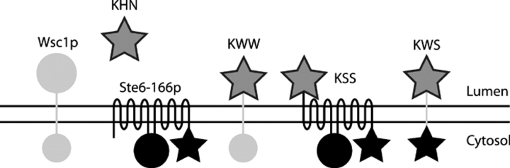
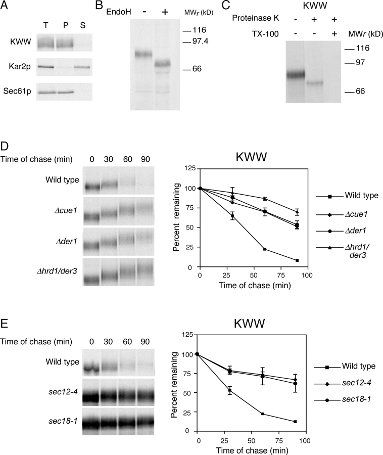
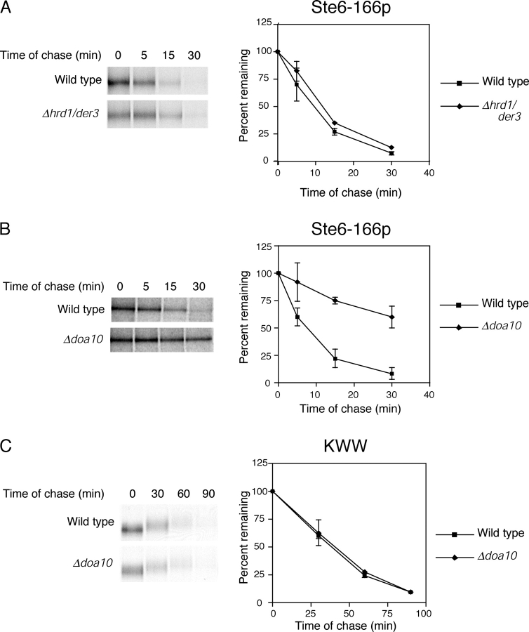
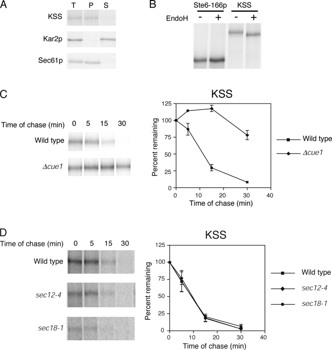
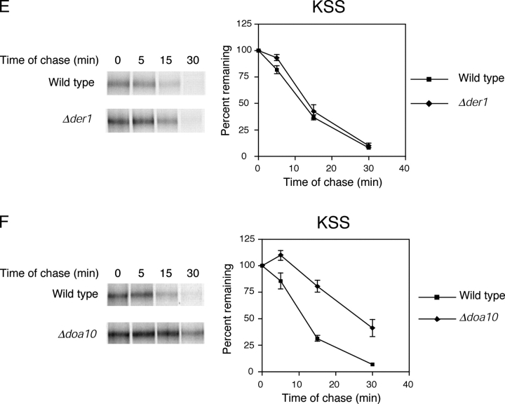
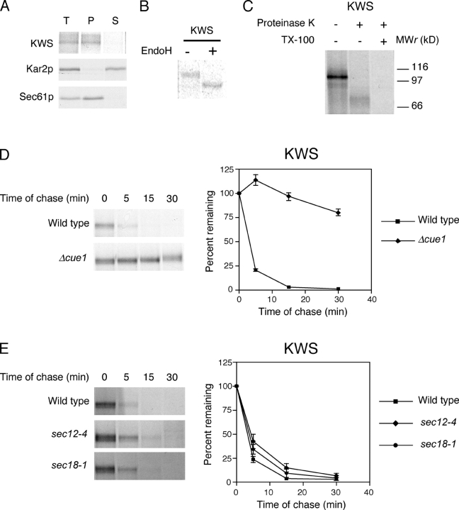
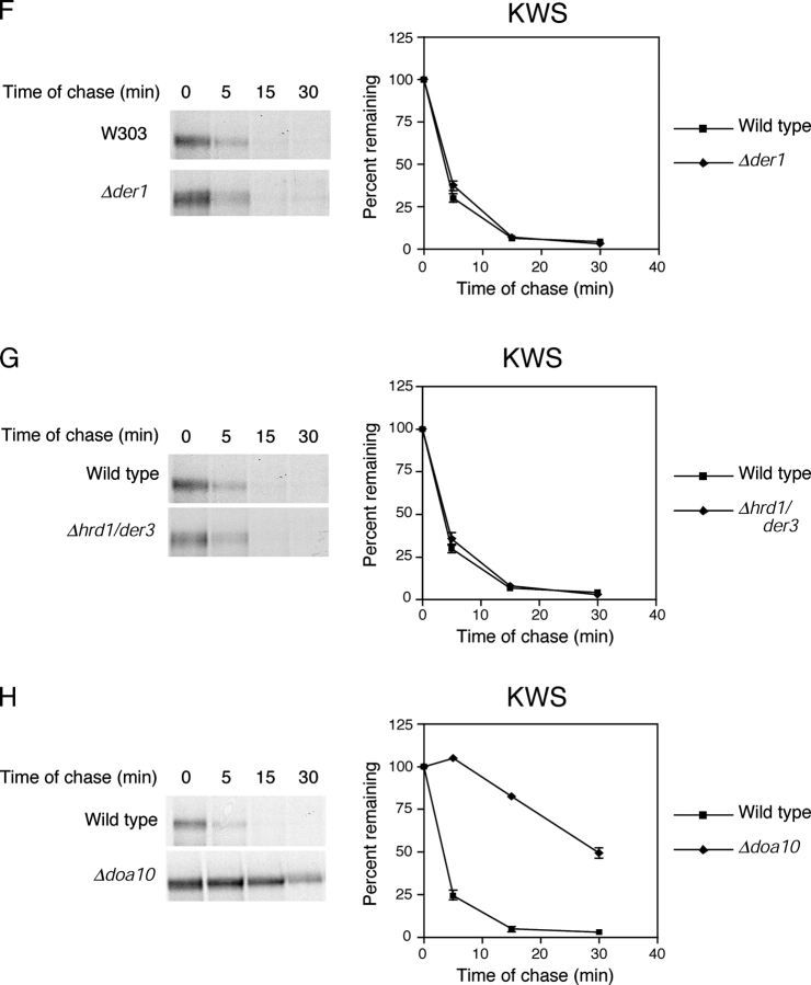
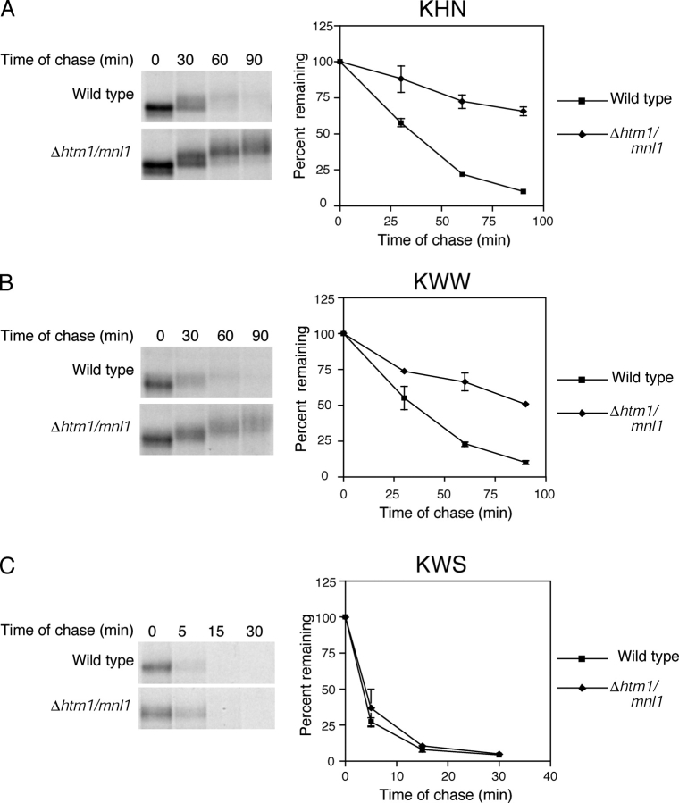
Misfolded proteins retained in the endoplasmic reticulum (ER) are degraded by the ER-associated degradation pathway. The mechanisms used to sort them from correctly folded proteins remain unclear. Analysis of substrates with defined folded and misfolded domains has revealed a system of sequential checkpoints that recognize topologically distinct domains of polypeptides. The first checkpoint examines the cytoplasmic domains of membrane proteins. If a lesion is detected, it is retained statically in the ER and rapidly degraded without regard to the state of its other domains. Proteins passing this test face a second checkpoint that monitors domains localized in the ER lumen. Proteins detected by this pathway are sorted from folded proteins and degraded by a quality control mechanism that requires ER-to-Golgi transport. Although the first checkpoint is obligatorily directed at membrane proteins, the second monitors both soluble and membrane proteins. Our data support a model whereby "properly folded" proteins are defined biologically as survivors that endure a series of distinct checkpoints.


Figures

Figure 1.
Figure 1.
Schematic representation of substrates. The engineered ERAD substrates bear three letter designations that describe their composition. The first, second, and third letters represent the luminal, transmembrane, and cytosolic domains, respectively. For each protein, KHN is symbolized by dark gray stars, Wsc1p portions by light gray bars (transmembrane) and circles (folded cytosolic and luminal domains), and Ste6-166p by black bars/loops (transmembrane), black circles (folded cytosolic domain), and black stars (misfolded cytosolic domain).
Figure 2.
Figure 2.
KWW is degraded by the ERAD-L pathway. (A) Cells expressing KWW were metabolically labeled with [35S]methionine/cysteine for 20 min. Cell lysates were prepared and incubated in 0.1 M of sodium carbonate, pH 11.0, for 30 min at 4°C and the membrane fraction separated by centrifugation. Proteins were immunoprecipitated from total (T), pellet (P), and supernatant (S) fractions. Sec61p and Kar2p serve as controls for integral membrane and soluble proteins, respectively. KWW was immunoprecipitated using anti-HA mAb (HA.11; Covance) and resolved by electrophoresis using 10% SDS polyacrylamide gels. (B) Cells expressing KWW were labeled with [35S]methionine/cysteine for 10 min. KWW immunoprecipitates were mock treated (−) or digested (+) with Endo H, resolved by SDS-PAGE, and visualized by autoradiography. (C) Protease protection assay. Wild-type cells expressing KWW were pulse labeled for 10 min and a fraction containing cytosol and membranes was prepared. The extracts were split and treated with proteinase K in the presence or absence of Triton X-100 or mock treated. KWW was immunoprecipitated and analyzed by SDS-PAGE (8%) and autoradiography. (D) Wild-type and mutant strains expressing KWW were pulse labeled for 10 min with [35S]methionine/cysteine followed by cold chase for times indicated. Immunoprecipitated proteins were analyzed by electrophoresis on 8% gels and visualized by autoradiography. (E) Wild-type, sec12-4, and sec18-1 strains were grown to log phase at 22°C and shifted to the restrictive temperature (37°C) for 30 min before pulse-chase analysis as described for D. The data were quantified by PhosphorImager analysis and plotted as mean values with SDs of two independent experiments (D and E). Representative autoradiograms are shown.
Figure 3.
Figure 3.
Ste6-166p requires the Doa10p E3 ubiquitin ligase for degradation. (A and B) Ste6-166p turnover was measured in wild-type, Δhrd1/der3, and Δdoa10 strains by pulse-chase assay as described in Fig. 2, except that cells were pulse labeled for 5 min followed by shorter times of chase. (C) The rate of KWW degradation was determined in wild-type and Δdoa10 cells as described in Fig. 2.
Figure 4.
Figure 4.
KSS is degraded by the ERAD-C pathway. (A) Carbonate alkali extraction assay was performed on wild-type cells expressing KSS as described in Fig. 2. (B) Cells expressing Ste6-166p or KSS were labeled with [35S]methionine/cysteine for 5 min and substrates immunoprecipitated from detergent lysates. N-linked carbohydrates were removed by Endo H digestion, proteins resolved by SDS-PAGE (7% gel), and visualized by autoradiography. (C) Wild-type and Δcue1 cells expressing KSS were labeled for 5 min with [35S]methionine/cysteine and chased for times indicated. KSS was resolved by electrophoresis using a 7% SDS polyacrylamide gel. (D) ER-to-Golgi transport is not required for degradation of KSS. Wild-type, sec12-4, and sec18-1 strains were grown to log phase at 22°C and then shifted to the restrictive temperature (37°C) for 30 min. (E and F) KSS turnover in Δder1 and Δdoa10 cells was performed as described for C.
Figure 4.
Figure 4.
KSS is degraded by the ERAD-C pathway. (A) Carbonate alkali extraction assay was performed on wild-type cells expressing KSS as described in Fig. 2. (B) Cells expressing Ste6-166p or KSS were labeled with [35S]methionine/cysteine for 5 min and substrates immunoprecipitated from detergent lysates. N-linked carbohydrates were removed by Endo H digestion, proteins resolved by SDS-PAGE (7% gel), and visualized by autoradiography. (C) Wild-type and Δcue1 cells expressing KSS were labeled for 5 min with [35S]methionine/cysteine and chased for times indicated. KSS was resolved by electrophoresis using a 7% SDS polyacrylamide gel. (D) ER-to-Golgi transport is not required for degradation of KSS. Wild-type, sec12-4, and sec18-1 strains were grown to log phase at 22°C and then shifted to the restrictive temperature (37°C) for 30 min. (E and F) KSS turnover in Δder1 and Δdoa10 cells was performed as described for C.
Figure 5.
Figure 5.
A misfolded cytosolic domain directs entry to the ERAD-C pathway. (A) Carbonate alkali extraction assay of KWS expressed in wild-type cells was performed as described in Fig. 2. T, total; P, pellet; S, supernatant. (B) KWS expressed in wild-type cells was pulse labeled for 5 min, immunoprecipitated, mock treated (−) and digested (+) with Endo H, and resolved by SDS-PAGE. (C) Protease protection assay was performed to determine the orientation of KWS in wild-type cells as described in Fig. 2, except that the proteins were resolved using a 10% polyacrylamide gel. (D–H) KWS stability was measured in wild-type and indicated mutant strains as described in Fig. 4.
Figure 5.
Figure 5.
A misfolded cytosolic domain directs entry to the ERAD-C pathway. (A) Carbonate alkali extraction assay of KWS expressed in wild-type cells was performed as described in Fig. 2. T, total; P, pellet; S, supernatant. (B) KWS expressed in wild-type cells was pulse labeled for 5 min, immunoprecipitated, mock treated (−) and digested (+) with Endo H, and resolved by SDS-PAGE. (C) Protease protection assay was performed to determine the orientation of KWS in wild-type cells as described in Fig. 2, except that the proteins were resolved using a 10% polyacrylamide gel. (D–H) KWS stability was measured in wild-type and indicated mutant strains as described in Fig. 4.
Figure 6.
Figure 6.
The Htm1p/Mnl1p ER lectin functions in the ERAD-L pathway. (A–C) Wild-type and Δhtm1/mnl1 mutant strains expressing KHN, KWW, or KWS were analyzed for substrate turnover as described for Fig. 4.

References

    1. Arvan, P., X. Zhao, J. Ramos-Castaneda, and A. Chang. 2002. Secretory pathway quality control operating in Golgi, plasmalemmal, and endosomal systems. Traffic. 3:771–780. - PubMed
    1. Barlowe, C., and R. Schekman. 1993. SEC12 encodes a guanine-nucleotide-exchange factor essential for transport vesicle budding from the ER. Nature. 365:347–349. - PubMed
    1. Bays, N.W., R.G. Gardner, L.P. Seelig, C.A. Joazeiro, and R.Y. Hampton. 2001. Hrd1p/Der3p is a membrane-anchored ubiquitin ligase required for ER- associated degradation. Nat. Cell Biol. 3:24–29. - PubMed
    1. Belden, W.J., and C. Barlowe. 2001. Role of Erv29p in collecting soluble secretory proteins into ER-derived transport vesicles. Science. 294:1528–1531. - PubMed
    1. Bonifacino, J.S., P. Cosson, N. Shah, and R.D. Klausner. 1991. Role of potentially charged transmembrane residues in targeting proteins for retention and degradation within the endoplasmic reticulum. EMBO J. 10:2783–2793. - PMC - PubMed

Publication types

MeSH terms