. 2004 May;78(9):4454-62.
doi: 10.1128/jvi.78.9.4454-4462.2004.

The human membrane cofactor CD46 is a receptor for species B adenovirus serotype 3

Affiliations

The human membrane cofactor CD46 is a receptor for species B adenovirus serotype 3

Dominique Sirena et al. J Virol. 2004 May.

Abstract

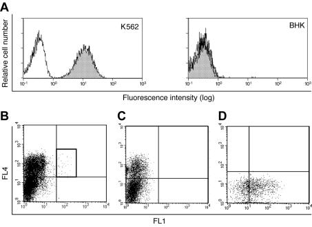
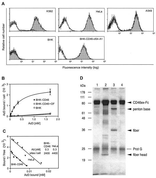
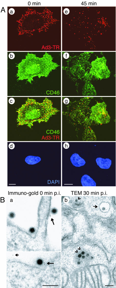
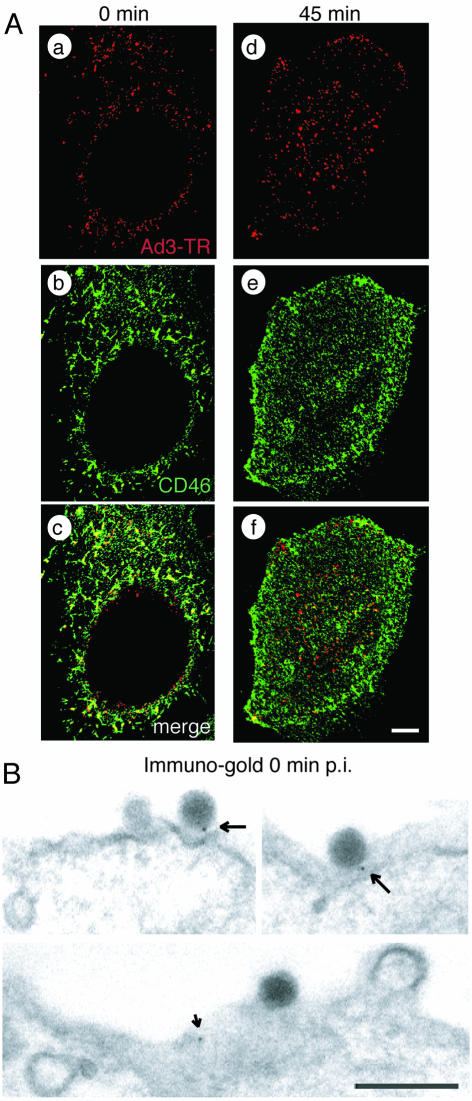
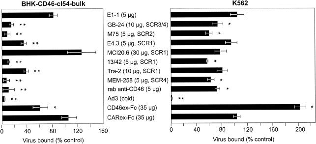
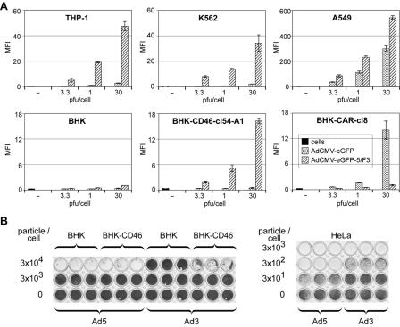
Many human adenovirus (Ad) serotypes use the coxsackie B virus-Ad receptor (CAR). Recently, CD46 was suggested to be a receptor of species B Ad serotype 11 (Ad11), Ad14, Ad16, Ad21, Ad35, and Ad50. Using Sindbis virus-mediated cDNA library expression, we identify here the membrane cofactor protein CD46 as a surface receptor of species B Ad3. All four major CD46 transcripts and one minor CD46 transcript expressed in nucleated human cells were isolated. Rodent BHK cells stably expressing the BC1 form of CD46 bound radiolabeled Ad3 with a dissociation constant of 0.3 nM, identical to that of CD46-positive HeLa cells expressing twice as many Ad3 binding sites. Pull-down experiments with recombinant Ad3 fibers and a soluble form of the CD46 extracellular domain linked to the Fc portion of human immunoglobulin G (CD46ex-Fc) indicated direct interactions of the Ad3 fiber knob with CD46ex-Fc but not CARex-Fc (Fc-linked extracellular domain of CAR). Ad3 colocalized with cell surface CD46 in both rodent and human cells at the light and electron microscopy levels. Anti-CD46 antibodies and CD46ex-Fc inhibited Ad3 binding to CD46-expressing BHK cells more than 10-fold and to human cells 2-fold. In CD46-expressing BHK cells, wild-type Ad3 and a chimeric Ad consisting of the Ad5 capsid and the Ad3 fiber elicited dose-dependent cytopathic effects and transgene expression, albeit less efficiently than in human cells. Together, our results show that all of the major splice forms of CD46 are predominant and functional binding sites of Ad3 on CD46-expressing rodent and human cells but may not be the sole receptor of species B Ads on human cells. These results have implications for understanding viral pathogenesis and therapeutic gene delivery.


Figures

FIG. 1.
FIG. 1.
CD46-expressing cells bind Ad3. (A) Flow cytometry of human leukemia K562 cells and rodent BHK cells that were not incubated with (white histograms) or that were incubated with (shaded histograms) Ad3- Alexa-488. (B and C) Single-cell sorting of BHK cells infected with Sindbis virus expressing a K562 cDNA library (B) and control Sindbis virus (C) after staining with propidium iodide (PI), anti-Sindbis virus antibodies (FL4), and Ad3- Alexa-488 (FL1). (D) PI-negative, Sindbis virus- and Ad3-positive cells (gate) were single cell sorted into 24-well plates containing BHK feeder cells and rescreened for Ad3- Alexa-488 binding. One of 30 positive clones is shown.
FIG. 2.
FIG. 2.
Ad3 binds with a high affinity to CD46-expressing cells through the fiber head. (A) Cytofluorometric analysis of CD46 expression levels in human and hamster cell lines. White histograms show staining with isotype control antibodies, and shaded histograms show CD46-specific staining. The cells used were human K562, HeLa, and A549 cells, parental BHK cells, and stably transfected BHK cells expressing the BC1 isoform of CD46. (B) Binding isotherms of Ad3 incubated with BHK-CD46-cl54 bulk cells or parental BHK cells. Nonspecific binding was determined in the presence of Ad3 DF. Error bar depicts standard error of the mean. (C) Scatchard analysis of Ad3 binding to BHK-CD46-cl54 bulk cells and HeLa cells. The number of binding sites per cell and the Kd values were calculated from the bound and the free virion concentrations determined by subtracting the bound virus from the input virus. (D) Direct interaction of the Ad3 fiber head with CD46ex-Fc. CD46ex-Fc was incubated without any addition (lane 1), with the purified fiber head (lane 2), with DF (lane 3), and with DP (lane 4). CD46ex-Fc was pulled down by protein G (Prot G)-Sepharose, and the SDS eluates were analyzed by SDS-PAGE and silver staining.
FIG. 3.
FIG. 3.
Ad3 colocalizes with cell surface CD46 of BHK-CD46-cl54-bulk cells. (A) CLSM analyses of TR-labeled Ad3 (red) and CD46 stained with non-function-blocking antibody MCI20.6 (green). Single sections were taken at 0 min (a to d) and at 5 min (e to h) p.i., including 4′,6′-diamidino-2-phenylindole (DAPI)-stained sections (d and h). Scale bars, 5 μm. (B) Transmission EM (TEM). (a) Immunogold staining of CD46 at 0 min p.i. Large arrows indicate Ad3 associated with protein A-gold directed to anti-CD46; the small arrow indicates a gold particle not associated with Ad3. (b) Ad3 internalization at 0 min p.i. Arrowheads indicate clusters of cytosolic Ad3; the small arrow indicates endosomal Ad3. Scale bars, 200 nm.
FIG. 4.
FIG. 4.
Ad3 colocalizes with cell surface CD46 of HeLa cells. (A) CLSM analyses of TR-labeled Ad3 (red) and CD46 stained with non-function-blocking antibody MCI20.6 (green). Single sections are shown at 0 min (a to c) and at 45 min (d to f) p.i. Scale bar, 5 μm. (B) Transmission EM showing immunogold staining of CD46 at 0 min p.i. Arrows indicate Ad3 associated with protein A-gold directed to anti-CD46; the small arrow indicates a gold particle not associated with Ad3. Scale bar, 200 nm.
FIG. 5.
FIG. 5.
CD46-dependent binding of Ad3 revealed by anti-CD46 antibodies and CD46ex-Fc. BHK-CD46-cl54 bulk cells and human K562 cells were incubated with the indicated antibodies, CD46ex-Fc, or CARex-Fc (control), followed by the addition of either Ad3- Alexa-488 or 3H-labeled Ad3. Virus binding was measured by flow cytometry or liquid scintillation counting. The asterisks indicate the level of significance (P values of <0.05 [single asterisks] and <0.005 [double asterisks] for comparisons with the negative controls E1-1 [anti-CAR] and CARex-Fc, respectively). rab, rabbit.
FIG. 6.
FIG. 6.
Transgene expression by chimeric Ad5/F3 and CPE of wild-type Ad3 in BHK-CD46 cells. (A) Ad-mediated eGFP expression in permissive human cells and CD46- or CAR-transfected rodent cells. The indicated cells were incubated with eGFP-expressing Ad5 or Ad5/F3 at different MOIs, and eGFP expression was analyzed at 2 days p.i. by flow cytometry. Results are shown as the mean fluorescence intensity (MFI). (B) CPE of wild-type Ad3 in CD46-transfected BHK cells and human HeLa cells and control infections of BHK-CD46 and HeLa cells with eGFP-expressing Ad5. Cells were infected with 10-fold dilutions of virus (3 × 101 to 3 × 10⁴ particles/cell) for 3 days, fixed with methanol, and stained with crystal violet.

References

    1. Adrian, T., G. Wadell, J. C. Hierholzer, and R. Wigand. 1986. DNA restriction analysis of adenovirus prototypes 1 to 41. Arch. Virol. 91:277-290. - PubMed
    1. Bergelson, J. M., J. A. Cunningham, G. Droguett, E. A. Kurt-Jones, A. Krithivas, J. S. Hong, M. S. Horwitz, R. L. Crowell, and R. W. Finberg. 1997. Isolation of a common receptor for coxsackie B viruses and adenoviruses 2 and 5. Science 275:1320-1323. - PubMed
    1. Casasnovas, J. M., M. Larvie, and T. Stehle. 1999. Crystal structure of two CD46 domains reveals an extended measles virus-binding surface. EMBO J. 18:2911-2922. - PMC - PubMed
    1. Chartier, C., E. Degryse, M. Gantzer, A. Dieterle, A. Pavirani, and M. Mehtali. 1996. Efficient generation of recombinant adenovirus vectors by homologous recombination in Escherichia coli. J. Virol. 70:4805-4810. - PMC - PubMed
    1. Chroboczek, J., R. W. H. Ruigrok, and S. Cusack. 1995. Adenovirus fiber, vol. I. Springer-Verlag KG, Berlin, Germany. - PubMed

Publication types

MeSH terms

LinkOut - more resources