. 2004 May;78(9):4838-46.
doi: 10.1128/jvi.78.9.4838-4846.2004.

In vitro replication of hepatitis E virus (HEV) genomes and of an HEV replicon expressing green fluorescent protein

Affiliations

In vitro replication of hepatitis E virus (HEV) genomes and of an HEV replicon expressing green fluorescent protein

Suzanne U Emerson et al. J Virol. 2004 May.

Abstract

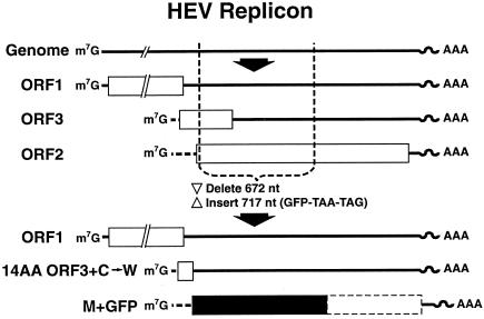
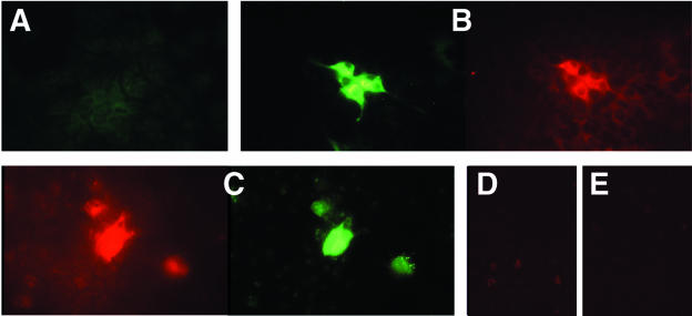
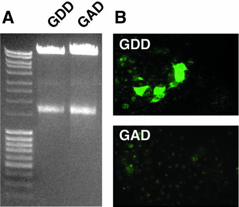
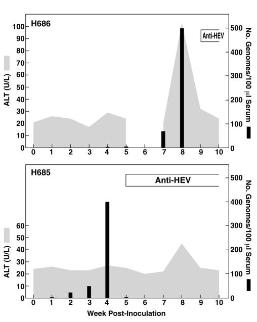
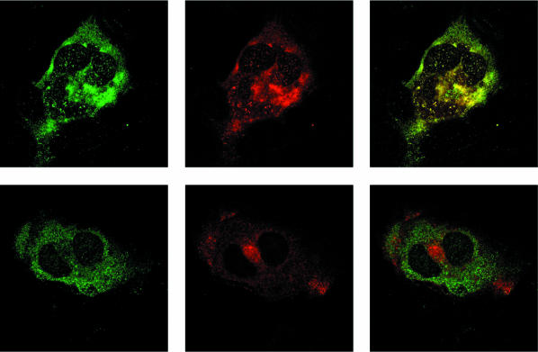
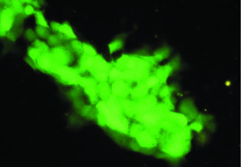
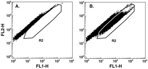
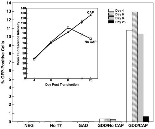
Hepatitis E virus (HEV) RNA replication occurred in seven of nine primate cell cultures transfected with in vitro transcripts of an infectious cDNA clone. Cell-to-cell spread did not occur in cell cultures, but rhesus monkeys inoculated with lysates of HEV-transfected PLC/PRF/5 and Huh-7 cells became infected with HEV. A replicon with the ORF2 and ORF3 genes deleted and replaced with the green fluorescent protein gene also replicated in the same primate cells that supported the replication of the full-length genome. Fluorescence-activated cell sorter analysis confirmed that the 7mG cap structure was critical for efficient infectivity, although replication could be initiated at a very low level in its absence. HEV virions were also able to infect a limited number of cells of certain lines.


Figures

FIG. 1.
FIG. 1.
Diagram of HEV replicon expressing GFP. The exact location of the 5′ end of each subgenomic RNA is unknown. Open rectangles, HEV coding regions; solid rectangle, GFP coding region; dashed-line rectangle, residual ORF2 gene (not translated). ORF1 is intact; ORF3 mRNA encodes the first 14 amino acids of ORF3 followed by a tryptophan and a termination codon; ORF2 mRNA encodes GFP followed by two termination codons.
FIG. 2.
FIG. 2.
Immune fluorescence microscopy of Huh-7 cells 14 days after transfection with the infectious HEV genome (A to C) or of mock-transfected Huh-7 cells (D and E). Cells were stained or costained with either chimpanzee 1313 preinfection serum (A), chimpanzee 1313 immune serum (green) and a monoclonal anti-ORF2 antibody (red) (B), a monoclonal anti-ORF3 antibody (red) and chimpanzee 1313 immune serum (green) (C), a monoclonal anti-ORF2 antibody (red) (D), or a monoclonal anti-ORF3 antibody (red) (E).
FIG. 3.
FIG. 3.
Transfection of cells with full-length genomes containing a wild-type (GDD) or a mutated (GAD) polymerase. (A) Agarose gel of 5 μl of each transcription reaction product used for transfection. (B) Immune fluorescence micrographs of representative cell monolayers stained with chimpanzee 1313 immune serum 14 days after transfection with wild-type (GDD) or mutated (GAD) genomes.
FIG. 4.
FIG. 4.
Course of HEV infection in rhesus macaques inoculated intravenously with cell lysates from transfected cell cultures. H686, lysate from PLC/PRF/5 cells. No sample was available for week 6. H685, lysate from Huh-7 cells. The ALT peak in the H685 graph was not at the correct time or high enough to be considered indicative of hepatitis.
FIG. 5.
FIG. 5.
Confocal microscopy of Huh-7 cells stained with anti-HEV at day 7 posttransfection. (Top left) Chimpanzee 1313 immune serum; (top center) anti-ORF2; (top right) merge. (Bottom left) Chimpanzee 1313 immune serum; (bottom center) anti-ORF3; (bottom right) merge. Magnification, ×126.
FIG. 6.
FIG. 6.
Fluorescence microscopy of Caco-2 cells producing GFP. Cell division has resulted in a colony containing more than 30 GFP-producing cells. Magnification, ×25.
FIG. 7.
FIG. 7.
Dot plot of FACS analysis. Cells, depicted as squares, are plotted as autofluorescence (FL2-H) versus fluorescence intensity in the FITC channel (FL1-H). (A) The pattern from control transfected cells was used to set the R2 window to exclude autofluorescent cells and to include cells containing GFP. (B) FACS distribution of cells 8 days after transfection with the GFP replicon. Only those cells that fell within the R2 area were scored as GFP positive.
FIG. 8.
FIG. 8.
FACS analysis of untransfected Huh-7 cells (NEG) or of Huh-7 cells transfected either with a transcription mixture from which polymerase had been omitted (No T7) or in which the GDD motif of the polymerase had been mutated to GAD (GAD) or with uncapped (GDD/No CAP) or capped (GDD/CAP) transcripts from the replicon containing the wild-type polymerase. A total of 100,000 cells were counted for each sample at each time. (Inset) Mean fluorescence intensity of GFP-positive cells from capped (circles) or uncapped (squares) sample.

References

    1. Agrawal, S., D. Gupta, and S. K. Panda. 2001. The 3′ end of hepatitis E virus (HEV) genome binds specifically to the viral RNA-dependent RNA polymerase (RdRp). Virology 282:87-101. - PubMed
    1. Arankalle, V. A., M. V. Joshi, A. M. Kulkarni, S. S. Gandhe, L. P. Chobe, S. S. Rautmare, A. C. Mishra, and V. S. Padbidri. 2001. Prevalence of anti-hepatitis E virus antibodies in different Indian animal species. J. Viral Hepat. 8:223-227. - PubMed
    1. Arankalle, V. A., S. A. Tsarev, M. S. Chadha, D. W. Alling, S. U. Emerson, K. Banerjee, and R. H. Purcell. 1995. Age-specific prevalence of antibodies to hepatitis A and E viruses in Pune, India, 1982 and 1992. J. Infect. Dis. 171:447-450. - PubMed
    1. Emerson, S. U., and R. H. Purcell. 2003. Hepatitis E virus. Rev. Med. Virol. 13:145-154. - PubMed
    1. Emerson, S. U., M. Zhang, X. J. Meng, H. Nguyen, M. St Claire, S. Govindarajan, Y. K. Huang, and R. H. Purcell. 2001. Recombinant hepatitis E virus genomes infectious for primates: importance of capping and discovery of a cis-reactive element. Proc. Natl. Acad. Sci. USA 98:15270-15275. - PMC - PubMed

Publication types

LinkOut - more resources