. 2004 May;78(9):4847-65.
doi: 10.1128/jvi.78.9.4847-4865.2004.

CCAAT/enhancer binding protein alpha binds to the Epstein-Barr virus (EBV) ZTA protein through oligomeric interactions and contributes to cooperative transcriptional activation of the ZTA promoter through direct binding to the ZII and ZIIIB motifs during induction of the EBV lytic cycle

Affiliations

CCAAT/enhancer binding protein alpha binds to the Epstein-Barr virus (EBV) ZTA protein through oligomeric interactions and contributes to cooperative transcriptional activation of the ZTA promoter through direct binding to the ZII and ZIIIB motifs during induction of the EBV lytic cycle

Frederick Y Wu et al. J Virol. 2004 May.

Abstract

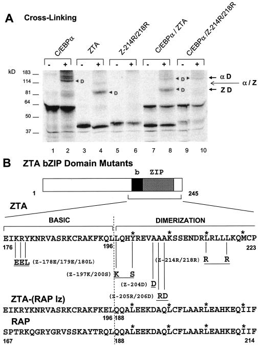
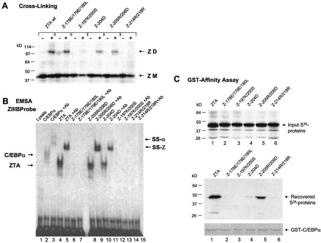
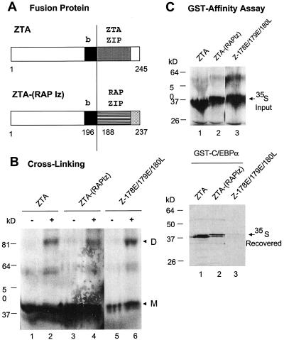
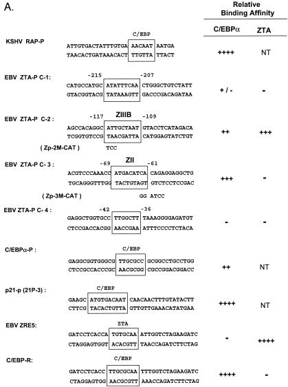
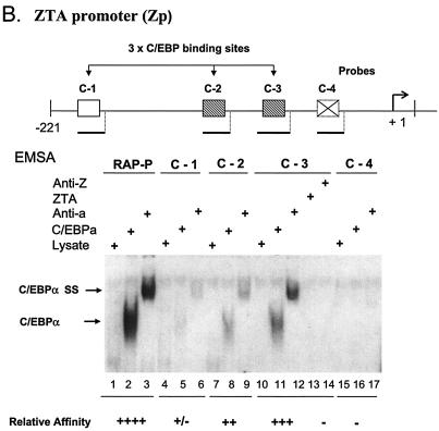
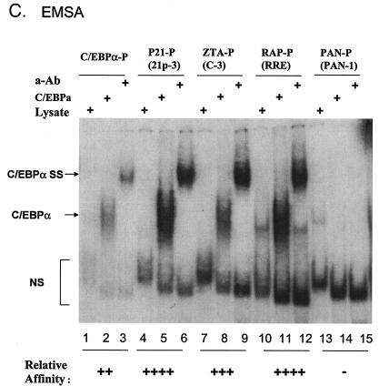
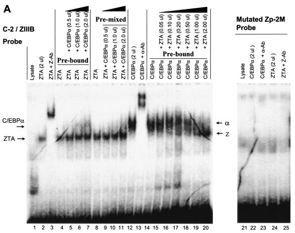
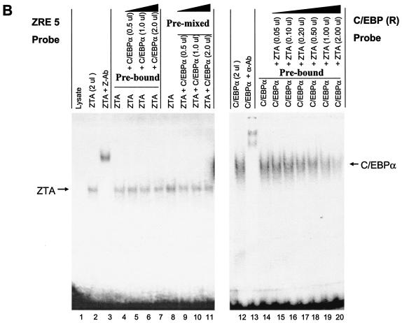
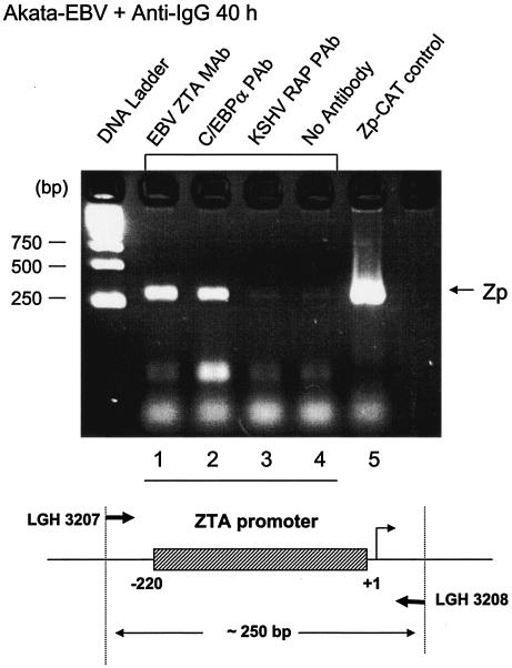
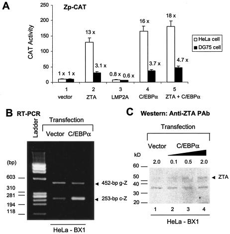
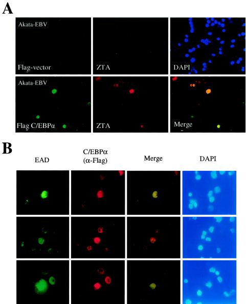
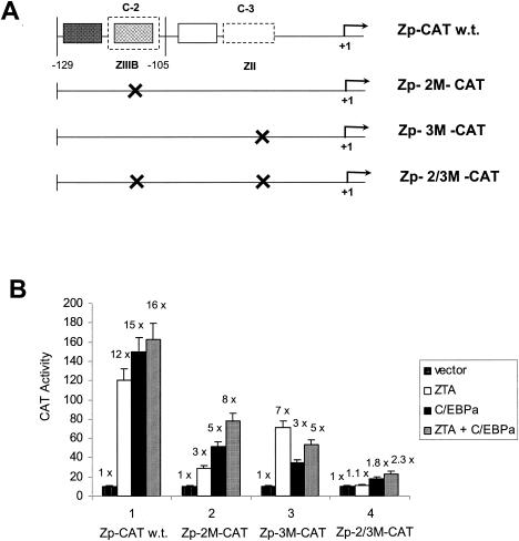
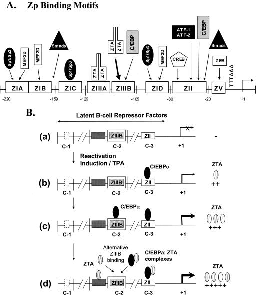
The Epstein-Barr virus (EBV)-encoded ZTA protein interacts strongly with and stabilizes the cellular CCAAT/enhancer binding protein alpha (C/EBPalpha), leading to the induction of p21-mediated G(1) cell cycle arrest. Despite the strong interaction between these two basic leucine zipper (bZIP) family proteins, the ZTA and C/EBPalpha subunits do not heterodimerize, as indicated by an in vitro cross-linking assay with in vitro-cotranslated (35)S-labeled C/EBPalpha and (35)S-labeled ZTA protein. Instead, they evidently form a higher-order oligomeric complex that competes with C/EBPalpha binding but not with ZTA binding in electrophoretic mobility shift assays (EMSAs). Glutathione S-transferase affinity assays with mutant ZTA proteins revealed that the basic DNA binding domain and the key leucine zipper residues required for homodimerization are all required for the interaction with C/EBPalpha. ZTA is known to bind to two ZRE sites within the ZTA promoter and to positively autoregulate its own expression in transient cotransfection assays, but there is conflicting evidence about whether it does so in vivo. Examination of the proximal ZTA upstream promoter region by in vitro EMSA analysis revealed two high-affinity C/EBP binding sites (C-2 and C-3), which overlap the ZII and ZIIIB motifs, implicated as playing a key role in lytic cycle induction. A chromatin immunoprecipitation assay confirmed the in vivo binding of both endogenous C/EBPalpha and ZTA protein to the ZTA promoter after lytic cycle induction but not during the latent state in EBV-infected Akata cells. Reporter assays revealed that cotransfected C/EBPalpha activated the ZTA promoter even more effectively than cotransfected ZTA. However, synergistic activation of the ZTA promoter was not observed when ZTA and C/EBPalpha were cotransfected together in either HeLa or DG75 cells. Mutagenesis of either the ZII or the ZIIIB sites in the ZTA promoter strongly reduced C/EBPalpha transactivation, suggesting that these sites act cooperatively. Furthermore, the introduction of exogenous C/EBPalpha into EBV-infected HeLa-BX1 cells induced endogenous ZTA mRNA and protein expression, as demonstrated by both reverse transcription-PCR and immunoblotting assays. Finally, double-label immunofluorescence assays suggested that EAD protein expression was activated even better than ZTA expression in latently infected C/EBPalpha-transfected Akata cells, perhaps because of the presence of a strong B-cell-specific repressed chromatin conformation on the ZTA promoter itself during EBV latency.


Figures

FIG. 1.
FIG. 1.
ZTA does not heterodimerize with C/EBPα. (A) Cross-linking of in vitro-translated 35S-labeled proteins with glutaraldehyde showing that C/EBPα and ZTA fail to form any novel heterodimer bands migrating at an intermediate position compared to the homodimer bands detected in control samples with each translated alone. Lanes: 1 and 2, C/EBPα from pYNC172a alone; 3 and 4, wild-type ZTA from pYNC100 alone; 5 and 6, mutant Z214R/218R from pFYW49 alone; 7 and 8, cotranslated C/EBPα and ZTA; 9 and 10, cotranslated C/EBPα and Z214R/218R. −, samples without cross-linking; +, samples with cross-linking. Arrowheads indicate dimers; αD, C/EBPα homodimers; ZD, ZTA homodimers; α/Z, expected position of C/EBPα-ZTA heterodimers. (B) Schematic diagram showing the specific amino acid changes in the relevant bZIP domain regions of the five ZTA mutants used in this study (upper sequence) and a comparison of the relevant amino acid sequences of the bZIP domain regions of wild-type ZTA, the ZTP-(RAP lz) fusion protein, and wild-type RAP. Asterisks indicate the expected leucine zipper positions.
FIG. 2.
FIG. 2.
Both the ZTA basic domain and the ability of ZTA to homodimerize are critical for the interaction with C/EBPα. (A) Cross-linking analysis of 35S-labeled in vitro-translated ZTA bZIP region point mutant proteins to determine their abilities to homodimerize in comparison with that of the wild-type (wt) ZTA protein. Only Z214R/218R from pFYW48 failed to homodimerize, and Z197K/200S from pFYW46 homodimerized at a lower efficiency. ZD, dimers; ZM, monomers. (B) EMSA showing the relative abilities of the five in vitro-translated ZTA bZIP region mutant proteins to bind to a 32P-labeled ZRE DNA probe (ZIIIB). Z178E/179E/180L (pFYW44), Z197K/200S (pFYW46), and Z214R/218R (pFYW49) all failed to bind to the ZIIIB site from the Zp. Positive controls included wild-type C/EBPα from pYNC172a (lanes 2 and 3) and wild-type ZTA from pYNC100 (lanes 4 and 5). Appropriate antibody (Ab) supershifts were carried out in the second lane of each pair to confirm that the shifted bands contained the expected protein. Arrows designate the positions of directly shifted bands (C/EBPα and ZTA) and antibody-supershifted complexes (SS-α and SS-Z, respectively). (C) GST affinity binding assays conducted with GST-C/EBPα to measure in vitro interaction affinities with the wild-type ZTA protein (lane 1) and each of the five ZTA bZIP region mutant proteins (lanes 2 to 6). (Upper panel) Input 35S-labeled in vitro-translated proteins. (Lower panels) 35S-labeled proteins recovered after binding to and elution from GST-C/EBPα beads (pSEW-C05). Only 35S-labeled wild-type ZTA from pYNC100 and 35S-labeled Z205R/206D from pFYW48 bound strongly to GST-C/EBPα; 35S-labeled Z204D bound at a lower efficiency. These results suggest that the basic domain as well as the ability of ZTA to homodimerize are both required for its interaction with C/EBPα.
FIG. 3.
FIG. 3.
A ZTA-(RAP lz) fusion protein still interacts with C/EBPα. (A) Schematic diagram showing the structures of wild-type ZTA and the ZTA-(RAP lz) fusion protein. b, ZTA basic domain; ZTA ZIP and RAP ZIP, C-terminal leucine zipper domains of ZTA and RAP, respectively. (B) Cross-linking of 35S-labeled in vitro-translated wild-type ZTA from pYNC100 (lanes 1 and 2), ZTA-(RAP lz) fusion fusion from pFYW04 (lanes 3 and 4), and the basic domain mutant Z178E/179E/180L from pFYW44 (lanes 5 and 6) demonstrating that the fusion protein retains the ability to homodimerize efficiently. −, samples without cross-linking; +, samples with cross-linking. D, dimers; M, monomers. (C) GST affinity assay showing that the ZTA-(RAP lz) fusion protein still binds efficiently to C/EBPα. (Upper panel) Input 35S-labeled in vitro-translated proteins. (Lower panel) Recovered bound 35S-labeled proteins eluted from GST-C/EBPα beads (pSEW-C05). Lanes: 1, wild-type ZTA (codons 1 to 245) from pYNC100; 2, ZTA-(RAP lz) fusion protein from pFYW04; 3, basic domain mutant ZTA178E/179E/180L from pFYW44.
FIG. 4.
FIG. 4.
Identification of two strong C/EBP binding sites in the EBV Zp. (A) DNA sequences of double-stranded oligonucleotide probes used in EMSA experiments and semiquantitive summary of their relative affinities for C/EBPα: +, ++, +++, and ++++, approximate values of one, two-, four-, and eightfold, respectively; NT, not tested. Known or projected C/EBPα recognition motifs are boxed. Several probes containing C/EBP sites that were evaluated previously (8) or that were recently identified in various cellular or viral promoters are also included (71, 72). (B) EMSA experiment with Zp-derived oligonucleotide probes showing that in vitro-translated C/EBPα from pYNC172a binds more strongly to the C-3 site (lanes 11 and 12) than to the C-2 site (lanes 8 and 9), only weakly to the more distal C-1 site (lanes 5 and 6), and not at all to the C-4 site (lanes 16 and 17). Wild-type ZTA from pYNC100 also failed to bind to the C-3 site (lanes 13 and 14). The relative positions of the four C site probes between positions −221 and +1 in the proximal Zp are indicated in the upper diagram. Anti-Z, added anti-ZTA PAb; Anti-α, added anti-C/EBPα PAb; SS, supershift. (C) EMSA experiment comparing the binding affinities of the Zp C-3 site and selected oligonucleotide probes containing various other known C/EBP sites. C/EBPα-P (lanes 1 to 3) is the C/EBP binding site from the C/EBPα promoter; P21-P (21p-3; lanes 4 to 6) is the strongest C/EBP binding site from the p21CIP-1 promoter (71); ZTA-P (C-3; lanes 7 to 9) is the C/EBP C-3 site from the Zp; RAP-P (RRE; lanes 10 to 12) is the C/EBP-II site from the KSHV RAP promoter (66); and PAN-P (PAN-1; lanes 13 to 15) is a negative control sequence taken from the KSHV PAN promoter, which contains no C/EBP sites. In each group, the first lane contains an unprogrammed reticulocyte lysate, the second lane contains in vitro-translated C/EBPα (pYNC172a), and the third lane contains both C/EBPα and added anti-C/EBPα PAb. NS, nonspecific binding.
FIG. 4.
FIG. 4.
Identification of two strong C/EBP binding sites in the EBV Zp. (A) DNA sequences of double-stranded oligonucleotide probes used in EMSA experiments and semiquantitive summary of their relative affinities for C/EBPα: +, ++, +++, and ++++, approximate values of one, two-, four-, and eightfold, respectively; NT, not tested. Known or projected C/EBPα recognition motifs are boxed. Several probes containing C/EBP sites that were evaluated previously (8) or that were recently identified in various cellular or viral promoters are also included (71, 72). (B) EMSA experiment with Zp-derived oligonucleotide probes showing that in vitro-translated C/EBPα from pYNC172a binds more strongly to the C-3 site (lanes 11 and 12) than to the C-2 site (lanes 8 and 9), only weakly to the more distal C-1 site (lanes 5 and 6), and not at all to the C-4 site (lanes 16 and 17). Wild-type ZTA from pYNC100 also failed to bind to the C-3 site (lanes 13 and 14). The relative positions of the four C site probes between positions −221 and +1 in the proximal Zp are indicated in the upper diagram. Anti-Z, added anti-ZTA PAb; Anti-α, added anti-C/EBPα PAb; SS, supershift. (C) EMSA experiment comparing the binding affinities of the Zp C-3 site and selected oligonucleotide probes containing various other known C/EBP sites. C/EBPα-P (lanes 1 to 3) is the C/EBP binding site from the C/EBPα promoter; P21-P (21p-3; lanes 4 to 6) is the strongest C/EBP binding site from the p21CIP-1 promoter (71); ZTA-P (C-3; lanes 7 to 9) is the C/EBP C-3 site from the Zp; RAP-P (RRE; lanes 10 to 12) is the C/EBP-II site from the KSHV RAP promoter (66); and PAN-P (PAN-1; lanes 13 to 15) is a negative control sequence taken from the KSHV PAN promoter, which contains no C/EBP sites. In each group, the first lane contains an unprogrammed reticulocyte lysate, the second lane contains in vitro-translated C/EBPα (pYNC172a), and the third lane contains both C/EBPα and added anti-C/EBPα PAb. NS, nonspecific binding.
FIG. 5.
FIG. 5.
ZTA interferes with C/EBPα binding to the C/EBP C-2/ZIIIB ZRE site, but C/EBPα has no effect on ZTA binding to the same site. (A) EMSA experiment showing the following results. First, in vitro-translated ZTA (pYNC100) and C/EBPα (pYNC172a) both bind strongly and independently to the 32P-labeled C-2 probe encompassing the ZIIIB ZRE motif (lanes 1, 2, 3, 12, and 13). Second, the addition of C/EBPα at various doses (lanes 4 to 11) does not affect ZTA DNA binding to the C-2 probe, whereas in the reciprocal approach, the addition of ZTA at various doses displaces C/EBPα binding to the C-2 probe (lanes 15 to 20). Third, point mutation of the C-2/ZIIIB site inactivates binding to both proteins (lanes 21 to 25). Z-Ab, anti-ZTA antibody; α-Ab, anti-C/EBPα antibody. (B) EMSA experiment showing the results of similar dose-response mixture binding experiments with in vitro-translated C/EBPα (pYNC172a) and ZTA (pYNC100) and with consensus 32P-labeled ZRE(5) and C/EBP(R) oligonucleotide DNA probes that each bind specifically to one protein but do not cross-react with the other (lanes 1, 2, 3, 12, and 13). Again, the addition of C/EBPα failed to affect the binding of ZTA to the ZRE(5) probe (lanes 4 to 11), but the addition of ZTA gradually displaced C/EBPα binding to the C/EBP(R) probe (lanes 14 to 20).
FIG. 5.
FIG. 5.
ZTA interferes with C/EBPα binding to the C/EBP C-2/ZIIIB ZRE site, but C/EBPα has no effect on ZTA binding to the same site. (A) EMSA experiment showing the following results. First, in vitro-translated ZTA (pYNC100) and C/EBPα (pYNC172a) both bind strongly and independently to the 32P-labeled C-2 probe encompassing the ZIIIB ZRE motif (lanes 1, 2, 3, 12, and 13). Second, the addition of C/EBPα at various doses (lanes 4 to 11) does not affect ZTA DNA binding to the C-2 probe, whereas in the reciprocal approach, the addition of ZTA at various doses displaces C/EBPα binding to the C-2 probe (lanes 15 to 20). Third, point mutation of the C-2/ZIIIB site inactivates binding to both proteins (lanes 21 to 25). Z-Ab, anti-ZTA antibody; α-Ab, anti-C/EBPα antibody. (B) EMSA experiment showing the results of similar dose-response mixture binding experiments with in vitro-translated C/EBPα (pYNC172a) and ZTA (pYNC100) and with consensus 32P-labeled ZRE(5) and C/EBP(R) oligonucleotide DNA probes that each bind specifically to one protein but do not cross-react with the other (lanes 1, 2, 3, 12, and 13). Again, the addition of C/EBPα failed to affect the binding of ZTA to the ZRE(5) probe (lanes 4 to 11), but the addition of ZTA gradually displaced C/EBPα binding to the C/EBP(R) probe (lanes 14 to 20).
FIG. 6.
FIG. 6.
ChIP assay showing that C/EBPα binds to Zp DNA in vivo. (Upper panel) ChIP assay results for EBV-positive Akata cells at 40 h after induction by anti-IgG treatment, showing that Zp DNA was recovered from both endogenous ZTA and C/EBPα immunoprecipitates but not from the negative control samples. Lanes contained Zp PCR DNA products obtained with an anti-ZTA MAb (1), an C/EBPα PAb (2), an anti-RAP PAb (3), or no antibody (4). Lane 5, positive control PCR amplification of a Zp-CAT plasmid (pHC41) with the same primers. (Lower panel) Schematic diagram of the Zp region (250 bp) that was targeted for detection by PCR amplification with specific primers LGH3207 and LGH3208.
FIG. 7.
FIG. 7.
Exogenous C/EBPα activates both a Zp reporter gene and endogenous ZTA mRNA and protein expression. (A) Transient reporter gene assays showing that C/EBPα activates the Zp-CAT promoter. (White bars) Target Zp (positions −200 to +10)-CAT reporter gene plasmid DNA (pHC41; 0.5 μg) was transfected into HeLa cells either alone or in the presence of cotransfected mammalian expression plasmids encoding empty vector DNA (pcLUC; 1.0 μg) (lane 1), ZTA (pRTS21; 0.1 μg) (lane 2), LMP2A (pLMP2A; 1.0 μg) (lane 3), C/EBPα (pSEW-C01; 1.0 μg) (lane 4), or a combination of ZTA and C/EBPα (lane 5). (Black bars) Assays were carried out with DG75 lymphocytes and the Zp-CAT reporter gene plasmid DNA (5 μg) electroporated either alone or together with expression plasmids encoding empty vector DNA (pcLUC; 10 μg), ZTA (pRTS21; 1 μg), LMP2A (pLMP2A; 10 μg), C/EBPα (pSEW-C01; 10 μg), or a combination of ZTA and C/EBPα. Total DNA was balanced by vector plasmid DNA as a carrier. Fold activation was calculated relative to the basal level of Zp-CAT activity in each cell type (designated 1.0). (B) Exogenous C/EBPα increases the level of ZTA mRNA in cells latently infected with EBV, as shown by the results of RT-PCR performed with mRNA harvested from C/EBPα-transfected HeLa-BX1 cells and primers specific for a spliced segment of the ZTA gene (LGH2617 and LGH2618). Relative to the basal level in HeLa-BX1 cells, the expression of ZTA mRNA (253-bp cDNA product; c-Z) was induced 10-fold in C/EBPα-transfected HeLa-BX1 cells. Some amplification of ZTA genomic DNA (452-bp band; g-Z) indicates equal loading. (C) Exogenous C/EBPα increases the level of ZTA protein in cells latently infected with EBV. Western blotting with an anti-ZTA MAb shows that the expression of C/EBPα from pSEW-C01 in HeLa-BX1 cells increased the total ZTA protein level by up to threefold in comparison to that in cells transfected with the same amount of the empty vector plasmid control.
FIG. 8.
FIG. 8.
Induction of endogenous ZTA and EAD protein expression by C/EBPα exogenously introduced into a B-lymphoblast cell line latently infected with EBV. (A) Introduction of Flag-C/EBPα encoded by pSEW-C02 into electroporated EBV-infected Akata cells. (Upper panel) Double-label IFA showing the lack of Flag-protein expression from empty vector control DNA, detected with an anti-Flag MAb (FITC, green) (left); the absence of enhanced ZTA protein expression in the same cell population, detected with an anti-ZTA PAb (rhodamine, red) (middle); and stained nuclear DNA in the whole-cell population (DAPI, blue) (right). (Lower panel) Double-label IFA showing the expression of Flag-C/EBPα from plasmid pSEW-C02, detected with an anti-Flag MAb (FITC, green) (left); the activation of ZTA protein expression in two out of four visible C/EBPα-positive cells, detected with an anti-ZTA PAb (rhodamine, red) (middle); and a merge of the two frames (yellow) (right). Induced ZTA expression was found to occur in 28% of Flag-C/EBPα-positive cells. (B) Activation of EBV EAD (PPF or BMLF1) protein expression by the introduction of exogenous Flag-C/EBPα plasmid DNA (pSEW-C02) into EBV-infected Akata cells by electroporation. All three sets of panels show examples of induced EAD expression detected by double-label IFA: EAD-expressing cells, detected with a mouse anti-EAD MAb (FITC, green) (first set of panels); C/EBPα-expressing cells, detected with a rabbit anti-Flag PAb (rhodamine, red) (second set of panels); a merge of those two sets of panels (yellow) (third set of panels); and staining of all nuclei in the same fields (DAPI, blue) (fourth set of panels). Induced EAD expression was found to occur in 85% of Flag-C/EBPα-positive cells.
FIG. 9.
FIG. 9.
Mutation of the C-3/ZII or C-2/ZIIIB C/EBP binding sites reduces or abolishes C/EBPα and ZTA transactivation of Zp-CAT. (A) Diagram showing the relative locations of the two strong C/EBP binding sites (C-2 and C-3) within the wild-type (w.t.) Zp-CAT target reporter gene and the structures of the three single- or double-point mutant Zp-CAT reporter genes used here. (B) Transient reporter gene assays in which effector plasmids encoding C/EBPα, ZTA, or both were cotransfected into HeLa cells with target Zp-CAT reporter plasmids containing either the intact Zp in Zp-CAT (pHC41) or its mutated derivatives Zp-2 M-CAT (pFYW33), Zp-3 M-CAT (pFYW42), and Zp-2/3 M-CAT (pFYW43). These results demonstrate that both the C-2/ZIIIB and the C-3/ZII sites are essential for high-level C/EBPα-mediated transactivation and that they both contribute to ZTA transactivation.
FIG. 10.
FIG. 10.
Updated summary and model of the known viral and cellular factors that bind to the Zp and play roles in regulating the switch from latency to the lytic cycle. (A) Simplified physical map of known multiple transcription factor and cellular repressor binding sites within the proximal segment of the Zp. (B) Schematic diagram hypothesizing a multistep role for C/EBPα at different stages of the initiation and ZTA-mediated amplification or positive autoregulation of Zp transactivation.

References

    1. Bauer, G., P. Hofler, and H. Zur Hausen. 1982. Epstein-Barr virus induction by a serum factor. I. Induction and cooperation with additional inducers. Virology 121:184-194. - PubMed
    1. Baumann, M., H. Mischak, S. Dammeier, W. Kolch, O. Gires, D. Pich, R. Zeidler, H. J. Delecluse, and W. Hammerschmidt. 1998. Activation of the Epstein-Barr virus transcription factor BZLF1 by 12-O-tetradecanoylphorbol-13-acetate-induced phosphorylation. J. Virol. 72:8105-8114. - PMC - PubMed
    1. Binne, U. K., W. Amon, and P. J. Farrell. 2002. Promoter sequences required for reactivation of Epstein-Barr virus from latency. J. Virol. 76:10282-10289. - PMC - PubMed
    1. Borras, A. M., L. Strominger, and S. H. Speck. 1996. Characterization of the ZI domains in the Epstein-Barr virus BZLF1 gene promoter: role in phorbol ester induction. J. Virol. 70:3894-3901. - PMC - PubMed
    1. Calkhoven, C. F., C. Muller, and A. Leutz. 2000. Translational control of C/EBPalpha and C/EBPbeta isoform expression. Genes Dev. 14:1920-1932. - PMC - PubMed

Publication types

MeSH terms

LinkOut - more resources