. 2004 Apr 20;101(16):6291-6.
doi: 10.1073/pnas.0401221101. Epub 2004 Apr 12.

Tobacco mosaic virus infection spreads cell to cell as intact replication complexes

Affiliations

Tobacco mosaic virus infection spreads cell to cell as intact replication complexes

Shigeki Kawakami et al. Proc Natl Acad Sci U S A. .

Abstract

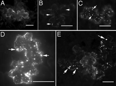
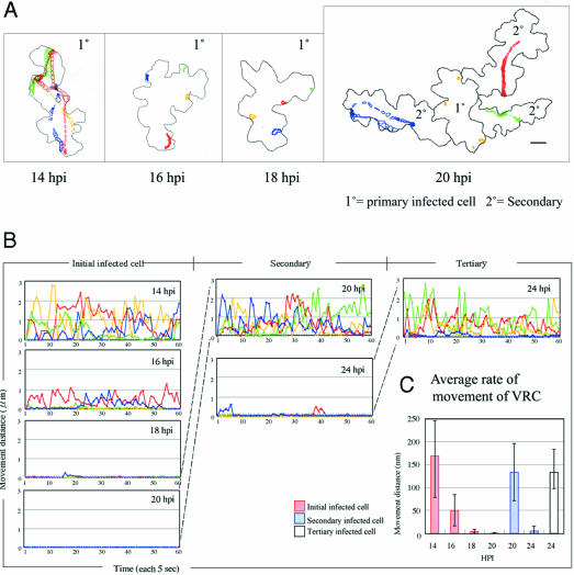
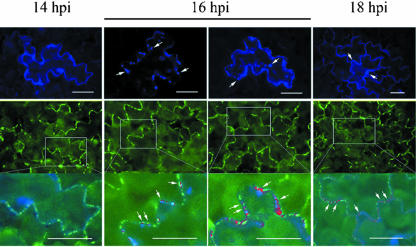
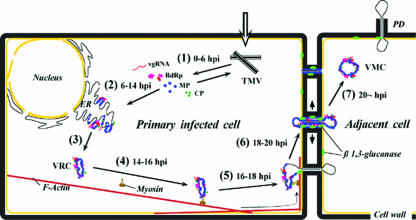
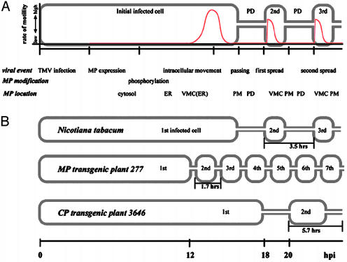
Plant viruses encode movement proteins (MPs) that facilitate cell-cell transport of infection through plasmodesmata. Intracellular and intercellular spread of virus replication complexes (VRCs) of tobacco mosaic virus was followed in intact leaf tissue from 12 to 36 h post infection (hpi) by using confocal microscopy. From 12 hpi, VRCs in primary infected cells were associated with cortical endoplasmic reticulum, and at 14 hpi, exhibited high intracellular mobility ( approximately 160 nm/sec); mobility was slowed between 14 and 16 hpi ( approximately 40 nm/sec), and by 18 hpi, VRCs were stationary, adjacent to plasmodesmata. VRCs traversed the plasmodesmata between 18 and 20 hpi. The process of formation and movement of VRCs was repeated in adjacent cells in 3-4 h vs. 20 h from primary infected cells. The rapid intracellular movement of the VRCs and the spread to adjacent cells was blocked by inhibitors of filamentous actin and myosin, but not by inhibitors of microtubules. We propose a model whereby cell-cell spread of tobamovirus infection is accomplished by subviral replication complexes that initiate TMV replication immediately after entry to adjacent cells.


Figures

Fig. 1.
Fig. 1.
Localization of MP:GFP produced by TMV infection in tobacco leaf tissue. After inoculation with transcripts of infectious clones, epidermal cells were observed by fluorescent microscopy at 14 (A and B), 16 (C), 18 (D), and 20 (E) hpi. Arrows indicate the VRCs. (Bars, 10 μm.)
Fig. 2.
Fig. 2.
Movement of VRCs in epidermal cells. (A) Tracing the movement of VRCs, showing compiled time-lapse images of infected epidermal cells. Movements of VRCs were traced in single infected cells for 5 min by using the nih image trace-object function to accumulate the sequential images (total = 60) of each VRC on the same frame. Four different colors show the movement of four independent VRCs. (Right) Four VRCs (yellow) in the primary infected cell do not move at 20 hpi, whereas VMCs in the secondary infected cells exhibit rapid intracellular movement. (Bars, 10 μm.) (B) Rate of movement of VRCs. The distance (μm) of VRC movement per 5-s interval was measured by nih image software (version 1.61); the distance functions of each of four VRCs were converted to rate graphs by Microsoft excel (version 98). The graphs show that the VRCs exhibit rapid intracellular movement in primary infected cells at 14 hpi; secondary cells at 20 hpi and tertiary infected cells at 24 hpi. Intercellular movement of the VRCs was halted on plasmodesmata of primary infected cells between 18 and 20 hpi and in secondary cells at 24 hpi. (C) Average rate of movement of VRCs. The graph presents the average of the intercellular movement of VRCs with SD at each time point (38) (See Movies 1–4).
Fig. 3.
Fig. 3.
Approach of VRCs and transmission of VMCs through plasmodesmata of MP:GFP(+)-transgenic plants. Leaves of plant line 266-A were infected with TMV expressing MP:eCFP and were examined by confocal microscopy. (Top) MP:eCFP (produced by infection; blue) in epidermal cells (visualized after excitation by 430 nm of light). (Middle) Location of MP:GFP produced by the transgene (visualized after excitation by 530 nm of light). (Bottom) Higher magnification of the plasmodesmata containing MP:GFP (green), virus MP:eCFP (blue), and colocalization (red) of GFP and eCFP. The study shows that in this plant line, MP:eCFP does not penetrate the plasmodesmata at 14 hpi, but does so at 16 hpi. After VRCs pass through the plasmodesmata to the secondary epidermal cells, both proteins can be colocalized in plasmodesmata or in the cytoplasm. In other cases, MP:GFP is pushed from the plasmodesmata and remains unassociated with MP:eCFP. The arrows indicate the VRCs (Top) and the colocalization (Bottom) of plant MP and VRCs. (Bars, 10 μm.)
Fig. 4.
Fig. 4.
Model of intercellular movement of VRCs in primary infected cell and cell–cell spread of VMCs. Step 1: from 0 to 6 hpi, TMV infection yields viral replicase (RdRp), MP, and CP, as well as viral RNA sequences. Step 2: from 6 to 14 hpi, viral RNA is produced as are viral proteins. At this time, MP is phosphorylated, and MP is localized on perinuclear and cytoplasmic ER to form VRCs. Steps 3 and 4: from 14 to 16 hpi, VRCs increase in size on cytoplasmic ER and in association with protein cytoskeleton, in particular with actin and myosin filaments. VRCs exhibit rapid intracellular movement in cytosol of infected cells. Step 5: from 16 to 18 hpi, movement of VRCs is stopped and some of the VRCs lodge, adjacent to plasmodesmata (PD). Step 6: from 18 to 20 hpi, plasmodesmata are modified and opened by action of the MP or VRCs, perhaps involving β-1,3 glucanase of the host (50, 51). Step 7: VMCs are spread from primary infected cells to adjacent cells, and replication is initiated in secondarily infected cells.
Fig. 5.
Fig. 5.
Timetable of TMV infection. (A) A timetable was produced on the basis of the results of observation of TMV MP:GFP virus infection in leaves of N. tabacum. Intracellular movement of VRCs in epidermal cells is represented by the red line. (B) Comparison of timetable of virus infection of TMV MP:GFP in transgenic plants. In plants that contains TMV MP (plant line 277), there was an increased rate of cell–cell spread, whereas plants that contain TMV CP (plant line 3646) exhibited reduced rate of spread of VMCs (Table 1).

References

    1. Carrington, J. C., Kasschau, K. D., Mahajan, S. K. & Schaad, M. C. (1996) Plant Cell 8, 1669-1681. - PMC - PubMed
    1. Beachy, R. N. & Heinlein, M. (2000) Traffic 1, 540-544. - PubMed
    1. Heinlein, M. (2002) Curr. Opin. Plant Biol. 5, 543-552. - PubMed
    1. Zambryski, P. (1995) Science 270, 1943-1944. - PubMed
    1. Robards, A. W. & Lucas, W. J. (1990) Annu. Rev. Plant Physiol. Plant Mol. Biol. 41, 369-419.

Publication types

LinkOut - more resources