. 2004 Apr 20;101(16):6021-6.
doi: 10.1073/pnas.0401590101. Epub 2004 Apr 12.

Regulation of spalt expression in the Drosophila wing blade in response to the Decapentaplegic signaling pathway

Affiliations

Regulation of spalt expression in the Drosophila wing blade in response to the Decapentaplegic signaling pathway

Rosa Barrio et al. Proc Natl Acad Sci U S A. .

Abstract

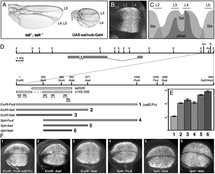
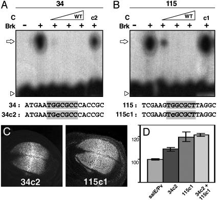
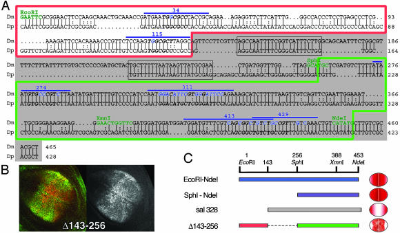
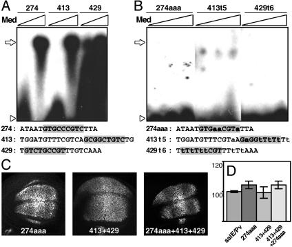
Pattern formation depends on the acquisition of precise cellular identities due to the differential expression of transcription factors. Enhancers within regulatory regions integrate the positive and negative regulatory signals directing gene transcription. Here, we analyze the enhancer that drives expression of the Drosophila gene spalt in the wing blade. This enhancer integrates positive signals, mediated by the Decapentaplegic signaling effector protein Medea, with the repressor activity of Brinker. The enhancer functions in the absence of binding sites for the wing-specific factor Scalloped. The molecular analysis of this enhancer indicates that there are additional factors yet unknown involved in the activation of spalt in the wing blade and that the mechanism of repression by Brinker does not rely on competition with Mad-Medea overlapping sites. The comparisons with other enhancers that respond to Decapentaplegic suggest that there are different possibilities to integrate the positive and negative inputs triggered by this signaling pathway.


Figures

Fig. 1.
Fig. 1.
Characterization of the sal wing blade-specific enhancer. (A) Phenotypic consequences of changes in sal and salr expression in adult wings. (Left) When both genes are absent (sal-, salr-), vein L2 does not form and L4 and L5 are fused. The intervein regions L2/3 and L4/5 disappear. (Right) When sal is expressed in the whole wing blade (UAS-sal/nub-Gal4), the veins L2 and L5 do not form, and the size of the wing is reduced. (B) Immunostaining showing Sal expression in a wild-type third-instar wing imaginal disc. Vein L2 forms in the anterior compartment in the region, where sal is expressed at low levels. The high levels of Sal coincide with the presumptive veins L3 and L4. In the posterior compartment, sal expression is adjacent to the presumptive vein L5. (C) Schematic representation of the antero/posterior axis of the wing blade. Phosphorylated Mad (pMad) colocalizes with high levels of Sal protein in the presumptive veins L3 and L4. pMad and sal are less abundant at the antero/posterior border. Brk overlaps with the regions of low levels of expression of sal. (D) Genomic region surrounding the sal transcript (gray boxes) according to Kühnlein et al. (21). Below, close-up of the 1.8-kb DNA fragment that contains the sal wing blade enhancer, indicating the relevant restriction enzymes used in our analysis. The oligonucleotides used in EMSA experiments are shown below, as well as the constructs sal328 and salE/Pv-Δ143–256. In the lower part, gray bars indicate the DNA fragments cloned in front of the reporter gene lacZ. EcoRI–PvuII corresponds to the salE/Pv enhancer. (E) Measurements of the areas of β-gal expression relative to the endogenous sal expression domain. Gray tones and numbers are according to D. Bars represent mean values and standard errors. Eliminating the EcoRI–SphI fragment increases the β-gal area by 21%. Eliminating the AseI–PvuII region also increases the β-gal area by 20%. (F) Third-instar wild-type wing imaginal discs stained with anti-Sal and anti-β-gal antibodies. The pictures show only the anti-β-gal single channels. Abbreviations for restriction enzymes: E, EcoRI; Pv, PvuII; R, RsaI; S, SalI; Xm, XmnI.
Fig. 2.
Fig. 2.
Brk is responsible for the repression of salE/Pv in the lateral regions of the wing blade. (A and B) EMSA experiments using radioactive oligonucleotides and bacterially expressed Brk protein at increasing concentrations. The sequences of the oligonucleotides used as probes and competitors are indicated under each panel. The Brk consensus binding sites are shaded in gray. Nucleotides mutated are indicated in lowercase. An open arrow indicates changes in mobility, whereas an arrowhead indicates the free probes. (A) Oligonucleotide 34 labeled radioactively shifts in the presence of recombinant Brk (+). The binding is competed for by increasing quantities of unlabeled wild-type oligonucleotide 34 (C). The mutant oligonucleotide 34c2 competes with the binding less efficiently (c2). (B) Oligonucleotide 115 binds recombinant Brk (+). Wild-type cold oligonucleotide 115 competes for the binding (C), whereas mutant 115c1 does not (c1). (C) β-gal expression driven by mutant salE/Pv enhancers in third-instar imaginal discs. Sal expression in the same discs is not shown. (D) Measurements of the areas of β-gal expression in relation to the endogenous sal expression. Note that mutation 115c1 increases β-gal expression area by 21%, whereas mutation 34c2 has a milder effect. When both sites are mutated, the reporter expression area increases similarly to single 155c1 mutant.
Fig. 5.
Fig. 5.
salE/Pv enhancer is conserved in D. pseudoobscura. (A) Alignment of D. melanogaster (Dm) and D. pseudoobscura (Dp) salE/Pv enhancers. Only the EcoRI–NdeI region of the enhancer is shown. Relevant restriction enzymes are shown in green. Oligonucleotides used in this analysis are indicated with a blue line. The nucleotides mutated in the different experiments are shown in blue. The red line boxes the repressor region, and the green line boxes the region important for activation. sal328 fragment is shaded in gray. The conserved regions eliminated in the mutant salE/Pv-Δ143–256 are boxed in gray. Italics and bold characters indicate consensus sequences for Brk, Med, or Sd. (B) Expression of Sal (green) and β-gal (red) in third-instar wing imaginal discs driven by the salE/Pv-Δ143–256 fragment. The corresponding red channel is in black and white. (C) Schematic representation of the EcoRI–NdeI fragment, which drives the reporter expression in the central part of the wing blade. When the EcoRI–SphI fragment is removed, the expression expands laterally (SphI–NdeI). The normal domain of expression is recovered when the repressor sites are added, albeit the levels of expression are heterogeneous (Δ143–256). However, the elimination of the repressor sites produces a change in the specificity of the enhancer that now drives reporter expression only in the lateral regions of the wing blade (sal328).
Fig. 3.
Fig. 3.
Med contributes to salE/Pv enhancer expression in certain regions of the wing blade. (A and B) EMSA experiments using radioactive oligonucleotides and bacterially expressed Med protein at increasing concentrations. The sequences of the oligonucleotides used as probes are indicated under each panel. The sequence consensus for Mad–Med binding is shaded in gray. The mutated nucleotides are indicated in lowercase. Open arrows indicate Med binding, whereas open arrowheads indicate the free probe. (A) Med binds to oligonucleotides 274, 413, and 429. (B) Mutated oligonucleotides 274aaa, 413t5, and 429t6 do not bind Med. (C) Expression of β-gal in third-instar discs driven by mutant salE/Pv enhancers. (D) Measurements of the areas of β-gal expression in relation to endogenous sal expression. The antero/posterior extension of the β-gal domain in the different mutants does not change in respect to the salE/Pv control.
Fig. 4.
Fig. 4.
Sd–Vg complex is not essential for sal expression in the wing blade. (A) EMSA radiograms of bacterially expressed Sd protein. Wild-type oligonucleotide 311 shifts in the presence of increasing quantities of Sd. Binding is abolished by mutation 311aa or greatly diminished by mutation 311tt. Below the panel, the above oligonucleotide sequences are shown, as well as the sequence of the 311.8n mutant. The mutations are indicated in lowercase. The AT core from the TEA domain (TEF1, TEC1, ABAA) consensus sequences is shaded, and arrows indicate the Sd consensus binding sites. Open arrows indicate Sd binding, whereas open arrowheads indicate the free probe. (B) β-gal expression driven by mutant salE/Pv in third-instar imaginal discs. Sal expression in the same discs is not shown. (C) Expression of Sal (green) and Vg (red) in early third-instar wing imaginal discs with large territories mutant for vg. Red (C′) and green (C″) channels are shown in black and white. Despite the absence of Vg, Sal is expressed in the blade. Orthogonal sections below C′ show the remaining Vg-expressing cells (arrow). Around them, sal-expressing cells appear in a different plane (C″, arrowhead).

References

    1. Affolter, M., Marty, T., Vigano, M. A. & Jazwinska, A. (2001) EMBO J. 20, 3298-3305. - PMC - PubMed
    1. de Celis, J. F. (2003) BioEssays 25, 443-451. - PubMed
    1. Muller, B., Hartmann, B., Pyrowolakis, G., Affolter, M. & Basler, K. (2003) Cell 113, 221-233. - PubMed
    1. Kirkpatrick, H., Johnson, K. & Laughon, A. (2001) J. Biol. Chem. 276, 18216-18222. - PubMed
    1. Rushlow, C., Colosimo, P. F., Lin, M. C., Xu, M. & Kirov, N. (2001) Genes Dev. 15, 340-351. - PMC - PubMed

Publication types

LinkOut - more resources