. 2004 Apr 20;101(16):6092-7.
doi: 10.1073/pnas.0304860101. Epub 2004 Apr 12.

Dendritic cells cross-present HIV antigens from live as well as apoptotic infected CD4+ T lymphocytes

Affiliations

Dendritic cells cross-present HIV antigens from live as well as apoptotic infected CD4+ T lymphocytes

Concepción Marañón et al. Proc Natl Acad Sci U S A. .

Abstract

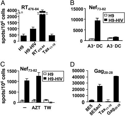
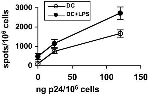
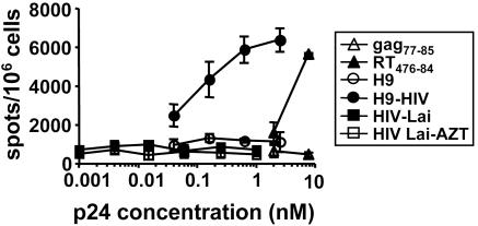
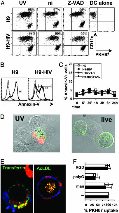
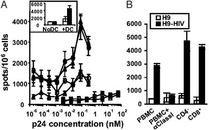
A better understanding of the antigen presentation pathways that lead to CD8(+) T cell recognition of HIV epitopes in vivo is needed to achieve better immune control of HIV replication. Here, we show that cross-presentation of very small amounts of HIV proteins from apoptotic infected CD4(+) T lymphocytes by dendritic cells to CD8(+) T cells is much more efficient than other known HIV presentation pathways, i.e., direct presentation of infectious virus or cross-presentation of defective virus. Unexpectedly, dendritic cells also take up actively antigens into endosomes from live infected CD4(+) T lymphocytes and cross-present them as efficiently as antigens derived from apoptotic infected cells. Moreover, live infected CD4(+) T cells costimulate cross-presenting dendritic cells in the process. Therefore, dendritic cells can present very small amounts of viral proteins from infected T cells either after apoptosis, which is frequent during HIV infection, or not. Thus, if HIV expression is transiently induced while costimulation is enhanced (for instance after IL-2 and IFNalpha immune therapy), this HIV antigen presentation pathway could be exploited to eradicate latently infected reservoirs, which are poorly recognized by patients' immune systems.


Figures

Fig. 1.
Fig. 1.
DC presentation of MHC class I molecule-restricted viral epitopes from HIV-infected apoptotic CD4+ T cells. (A) DC were incubated with apoptotic cells purified from irradiated H9 or H9-HIV cells at an H9:DC ratio of 3:1, then with LPS, and tested by using a CD8+ T cell line specific for RT476–84 epitope at a DC:effector ratio of 1:3. As control, DC were incubated with 1 μM peptide. (B) The test was carried out by using DC expressing or not the class I MHC restriction molecule HLA-A3. (C) DC:H9 coculture was performed in the presence or not of AZT or in transwells (TW). (D) DC were cultured with irradiated 8E5 cells expressing viral proteins after activation (8E5act) in the absence of LPS and tested as before. Data are representative of at least three experiments each, except for B and C (two experiments).
Fig. 2.
Fig. 2.
DC cross-present a viral epitope from CD4+ primary lymphocytes infected with a WT HIV isolate. DC were cultured with irradiated CD4+ T cell blasts infected with different amounts of a primary HIV isolate in the presence or absence of LPS and tested by using an anti-Nef73–82 CD8+ line as in Fig. 1. This experiment was carried out in the presence of AZT.
Fig. 3.
Fig. 3.
Efficiency of cross-presentation of HIV antigens from apoptotic cells compared with free virus. DC were cultured with different amounts and sources of HIV epitope: synthetic peptides, UV-irradiated, apoptotic H9 and H9-HIV cells in the presence of AZT, or HIV-1-Lai with or without AZT; then LPS was added. After coculture, DC were tested by using an anti-RT476–84 CD8+ line. Equivalent Gag p24 values were calculated after titration of viruses or lysed cells. For synthetic peptides, molar concentrations are represented. Data are representative of two experiments.
Fig. 4.
Fig. 4.
Uptake of cell material from HIV-infected live cells by DC. (A) CD11c and PKH67 labeling of DC gated using forward scatter (FSC) and side scatter (SSC) criteria (note: some H9 cells appear in this gate as CD11c-negative events) after overnight coculture with PKH67-labeled H9 cells at an H9:DC ratio of 3:1. Dot plots show DC that have acquired H9-derived material as CD11c+ PHK67+ events, and their percentages are noted. UV, UV-irradiated; ni, nonirradiated; Z-VAD, nonirradiated Z-VAD-treated H9 cells. (B) Annexin-V staining of H9 cells gated using FSC and SSC criteria and CD11c-, PKH67+ labeling after overnight incubation alone (dotted lines), or with DC (thick line, UV-irradiated; thin line, nonirradiated H9 cells). (C) Annexin-V staining of H9 cells after different incubation times with DC. (D) Microscopy visualization of DC interaction with H9-HIV cells. Transmission light and confocal fluorescence overlay. DC were cultured for 15 min with UV-irradiated (UV) or nonirradiated (live) H9-HIV cells, treated with Z-VAD. They were fixed, permeabilized, and labeled for CD3 (green) and TUNEL (terminal deoxynucleotidyltransferase-mediated dUTP nick end labeling) reaction (red) to detect apoptotic cells. Intimate membrane interactions were found between DC (unlabeled) and H9-HIV cells (green). (E) Material from live, infected, PKH26-labeled (red) CD4+ T cell blasts is internalized and colocalizes (in yellow) with transferrin or AcLDL after 2 h of incubation with DC. (F) Competition of live (filled) or UV-irradiated (open) H9-HIV-associated (positive for CD11c, in blue) PKH67 uptake by DC using several adhesion molecule ligands. man, mannan. Mean values and SDs of at least three independent experiments are represented. DC were 60–95% PKH67-positive in absence of competitors. Data are representative of at least three experiments, except for C (two experiments).
Fig. 5.
Fig. 5.
Cross-presentation of HIV antigens from live infected cells. (A) DC were tested as in Fig. 3 after coculture with UV-irradiated (circles) or live, Z-VAD-treated (triangles) H9 (empty symbols) or H9-HIV (filled symbols) cells in the presence of AZT, by using an anti-RT476–84 CD8+ line (DC:T ratio 1:1), all in the presence of LPS. DC were also incubated with free viral particles in the presence (open squares) or absence (filled squares) of AZT as in Fig. 3. (Inset) Live H9 (open bars) and H9-HIV (filled bars) cells were incubated or not with DC in the presence of Z-VAD and tested as before. (B) Recognition of DC cocultured with Z-VAD-treated H9-HIV cells by circulating lymphocytes from an HIV+ patient (DC:T ratio 1:5) in the presence of LPS and AZT. PBMC were incubated with an anti-class I ascitis, CD4-depleted or CD8-enriched. Data are representative of two experiments.

Similar articles

Cited by

References

    1. Letvin, N. L. & Walker, B. D. (2003) Nat. Med. 9, 861-866. - PubMed
    1. Wilson, J. D., Ogg, G. S., Allen, R. L., Davis, C., Shaunak, S., Downie, J., Dyer, W., Workman, C., Sullivan, S., McMichael, A. J. & Rowland-Jones, S. L. (2000) AIDS 14, 225-233. - PubMed
    1. Dalod, M., Dupuis, M., Deschemin, J.-C., Goujard, C., Deveau, C., Meyer, L., Ngo, N., Rouzioux, C., Guillet, J.-G., Delfraissy, J.-F., et al. (1999) J. Clin. Invest. 104, 1431-1439. - PMC - PubMed
    1. Blankson, J. N., Persaud, D. & Siliciano, R. F. (2002) Annu. Rev. Med. 53, 557-593. - PubMed
    1. Jung, S., Unutmaz, D., Wong, P., Sano, G., De los Santos, K., Sparwasser, T., Wu, S., Vuthoori, S., Ko, K., Zavala, F., et al. (2002) Immunity 17, 211-220. - PMC - PubMed

Publication types