. 2004 Apr 20;101(16):5910-5.
doi: 10.1073/pnas.0400891101. Epub 2004 Apr 12.

Molecular basis for the inhibition of the carboxyltransferase domain of acetyl-coenzyme-A carboxylase by haloxyfop and diclofop

Affiliations

Molecular basis for the inhibition of the carboxyltransferase domain of acetyl-coenzyme-A carboxylase by haloxyfop and diclofop

Hailong Zhang et al. Proc Natl Acad Sci U S A. .

Abstract

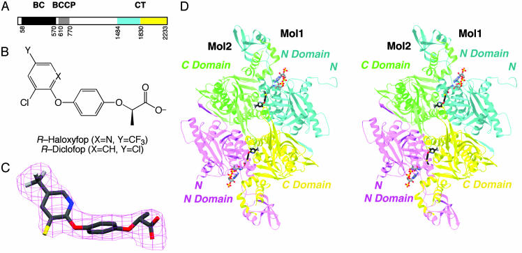
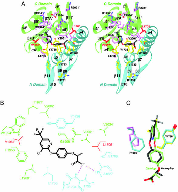
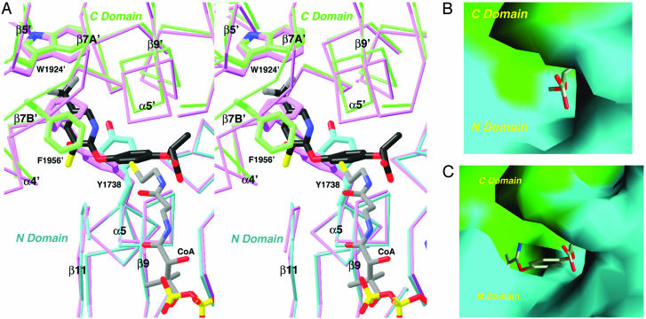
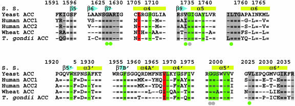
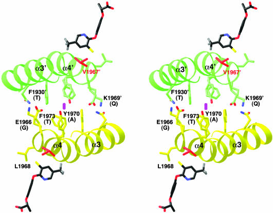
Acetyl-CoA carboxylases (ACCs) are crucial for the metabolism of fatty acids, making these enzymes important targets for the development of therapeutics against obesity, diabetes, and other diseases. The carboxyltransferase (CT) domain of ACC is the site of action of commercial herbicides, such as haloxyfop, diclofop, and sethoxydim. We have determined the crystal structures at up to 2.5-A resolution of the CT domain of yeast ACC in complex with the herbicide haloxyfop or diclofop. The inhibitors are bound in the active site, at the interface of the dimer of the CT domain. Unexpectedly, inhibitor binding requires large conformational changes for several residues in this interface, which create a highly conserved hydrophobic pocket that extends deeply into the core of the dimer. Two residues that affect herbicide sensitivity are located in this binding site, and mutation of these residues disrupts the structure of the domain. Other residues in the binding site are strictly conserved among the CT domains.


Figures

Fig. 1.
Fig. 1.
Crystal structure of CT domain in complex with haloxyfop. (A) Domain organization of yeast ACC. The N and C subdomains of CT are colored in cyan and yellow, respectively. (B) Chemical structures of the herbicides (R)-haloxyfop and (R)-diclofop. (C) Final 2FoFc electron density at 2.8-Å resolution for haloxyfop, contoured at 1σ. (D) Schematic stereodrawing of the structure of yeast CT domain dimer in complex with haloxyfop. The N domains of the two monomers are colored in cyan and magenta, and the C domains are colored in yellow and green. The inhibitor is shown in stick models, in black for carbon atoms. The CoA molecule is shown for reference (11), in gray. C was produced with setor (28), and D was produced with ribbons (29).
Fig. 2.
Fig. 2.
The binding mode of haloxyfop. (A) Stereographic drawing showing the binding site for haloxyfop. The N domain of one monomer is colored in cyan, and the C domain of the other monomer is in green. The side chains of residues in the binding site are shown in yellow and magenta, respectively. The dashed segment indicates the disordered residues 1959′–1964′. The drawing was produced with ribbons (29). (B) Schematic drawing of the interactions between haloxyfop and the CT domain. (C) Overlay of the binding mode of haloxyfop (in black) and diclofop (in green). The conformations of residues Tyr-1738 and Phe-1956′ in the haloxyfop (yellow and magenta) and diclofop (cyan) complexes are also shown.
Fig. 3.
Fig. 3.
Conformational change in the CT domain upon inhibitor binding. (A) Stereographic structural overlay of the CT domain free enzyme (in magenta) and the haloxyfop complex (in cyan and green for the N and C domains) near the inhibitor binding site. The binding mode of CoA (11) is also shown. The poorer structural overlap in the C domain is due to the change in the dimer organization. (B) Molecular surface of the active site of the free enzyme. The model of haloxyfop is included for reference. Most of the inhibitor is in steric clash with the enzyme. (C) Molecular surface of the binding site in the haloxyfop complex. For both B and C, residues 1759–1772 and 2026′–2098′ have been removed to give a better view of the binding site. A was produced with ribbons (29), and B and C were produced with grasp (30).
Fig. 4.
Fig. 4.
Sequence conservation in the binding site. Sequence alignment of residues in the haloxyfop (in green) and CoA (in gray) binding pockets. The two residues that confer herbicide resistance, Leu-1705 and Val-1967, are highlighted in red. A dash represents a residue that is identical to that in yeast ACC, whereas an equals sign represents a residue that is strictly conserved among ACCs. S. S., secondary structure.
Fig. 5.
Fig. 5.
Differences between yeast and plant ACCs in the dimer interface of the CT domain. In this stereographic drawing, the α3 and α4 helices of one monomer are shown in yellow, and those of the other monomer are shown in green. The side chains in the dimer interface are shown and labeled. The equivalent residues in the plant ACCs are shown in parentheses. The haloxyfop molecules are shown for reference. The twofold axis of the dimer is indicated by the magenta oval.

Similar articles

Cited by

References

    1. Friedman, J. M. (2003) Science 299, 856–858. - PubMed
    1. Hill, J. O., Wyatt, H. R., Reed, G. W. & Peters, J. C. (2003) Science 299, 853–855. - PubMed
    1. Pi-Sunyer, X. (2003) Science 299, 859–860. - PubMed
    1. Alberts, A. W. & Vagelos, P. R. (1972) in The Enzymes, ed. Boyer, P. D. (Academic, New York), Vol. 6, pp. 37–82.
    1. Wakil, S. J., Stoops, J. K. & Joshi, V. C. (1983) Annu. Rev. Biochem. 52, 537–579. - PubMed

Publication types

MeSH terms