

Increased transendothelial migration of scleroderma lymphocytes
- PMID: 15082489
- PMCID: PMC1754998
- DOI: 10.1136/ard.2002.004838
Increased transendothelial migration of scleroderma lymphocytes
Abstract
Background: CD4+ T lymphocytes play an important part in the pathogenesis of scleroderma (systemic sclerosis, SSc) and predominate in perivascular SSc skin lesions. Both soluble and membrane bound adhesion molecules are overexpressed in SSc, possibly influencing lymphocyte/endothelial cell (EC) contact.
Objective: To assess the transendothelial migration capacity of peripheral lymphocytes in vitro.
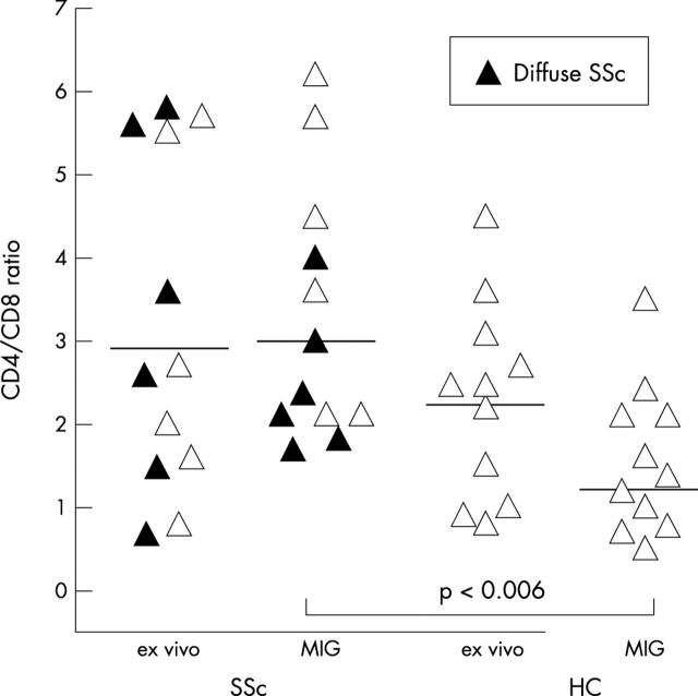
Patients and methods: Collagen was covered with human umbilical vein endothelial cells (HUVEC), and peripheral blood mononuclear cells (PBMC) of patients and matched healthy controls (HC) were added in parallel experiments. Before and after fractionated harvest of non-adherent, bound, and migrated lymphocytes, the CD4/CD8 ratio and the lymphocytic expression of activation markers and adhesion molecules were analysed by fluorocytometry.
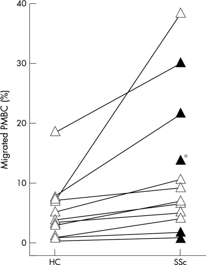
Results: 13 (SD 12)% of the SSc PBMC migrated compared with only 5 (5)% HC PBMC (p<0.0002); this increase was primarily due to the migration of CD3+ T lymphocytes and mainly to a larger proportion of CD4+ cells within this CD3+ fraction (71 (SD 14)% for SSc v 56 (14)% for HC, p<0.03), leading to an increased CD4/CD8 ratio among migrated SSc lymphocytes in comparison with controls (3.3 (1.5) v 1.62 (0.93), p<0.006). Among migrated SSc CD4+ T lymphocytes, the frequency of HLA-DR+ cells was increased; migrated lymphocytes highly expressed the adhesion molecules CD11a, CD49d, CD29, and CD44.
Conclusion: Transendothelial migration of CD4+ T lymphocytes is enhanced in SSc, and migrating cells exhibit an activated phenotype. The data suggest that activated CD3+CD4+ lymphocytes as found in SSc peripheral blood are prone to transvascular migration, thus contributing to the formation of typical perivascular lymphocytic infiltrates.
Figures

References
MeSH terms
Substances
LinkOut - more resources
Full Text Sources
Medical
Research Materials
Miscellaneous
