. 2004 May;24(9):3577-87.
doi: 10.1128/MCB.24.9.3577-3587.2004.

The Apc5 subunit of the anaphase-promoting complex/cyclosome interacts with poly(A) binding protein and represses internal ribosome entry site-mediated translation

Affiliations

The Apc5 subunit of the anaphase-promoting complex/cyclosome interacts with poly(A) binding protein and represses internal ribosome entry site-mediated translation

Nadejda Koloteva-Levine et al. Mol Cell Biol. 2004 May.

Abstract

The anaphase-promoting complex/cyclosome (APC/C) is a multisubunit ubiquitin ligase that mediates the proteolysis of cell cycle proteins in mitosis and G(1). We used a yeast three-hybrid screen to identify proteins that interact with the internal ribosome entry site (IRES) of platelet-derived growth factor 2 mRNA. Surprisingly, this screen identified Apc5, although it does not harbor a classical RNA binding domain. We found that Apc5 binds the poly(A) binding protein (PABP), which directly binds the IRES element. PABP was found to enhance IRES-mediated translation, whereas Apc5 overexpression counteracted this effect. In addition to its association with the APC/C complex, Apc5 binds much heavier complexes and cosediments with the ribosomal fraction. In contrast to Apc3, which is associated only with the APC/C and remains intact during differentiation, Apc5 is degraded upon megakaryocytic differentiation in correlation with IRES activation. Expression of Apc5 in differentiated cells abolished IRES activation. This is the first report implying an additional role for an APC/C subunit, apart from its being part of the APC/C complex.


Figures
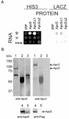
FIG. 1.
FIG. 1.
(A) The C-terminal half of Apc5 promotes the generation of a stable three-hybrid complex with IRES RNA. pACTII-IRP, pACTII-Apc5, pACTII-Apc5Δ1, or pACTII-Apc5Δ2 expressing IRP, full-length Apc5, or the C- or N-terminal part of Apc5, respectively, were cotransformed along with pDBRevM10-B, pDBRevM10-A, or pDBRevM10 expressing the different RNA baits into CG1945 and Y190 yeast strains. Colonies were selected for HIS3 expression and assayed for LacZ expression as described in Materials and Methods. (B) Total extract from K562 cells was immunoprecipitated by anti-Apc3 antibodies (lanes 1) or a large excess of anti-HA antibodies (lanes 2) or was incubated with beads only (lanes 3). HEK293 cells were infected with vTF7-3 and transfected with pFlag-Apc5 (lanes 4) or pCDNA3-Apc5 (lanes 5). The membranes were immunoblotted with antibodies specific for Apc5, Apc3, or Flag tag, as indicated at the bottom of each panel.
FIG. 2.
FIG. 2.
Apc5 interacts with PABP. (A) Yeast two-hybrid system. pGBT9-Apc5(395-755) expressing the C-terminal half of human Apc5 fused to the GAL4 DNA binding domain was cotransfected into CG1945 yeast strain along with pACTII-hPABP, pGAD424-yPab1(1-237), pGAD424-yPab1(189-346), pGAD424-yPab1(345-577), or pACTII-IRP expressing the human PABP, different regions of yeast Pab1, or IRP, respectively, fused to the GAL4 activation domain. Colonies were selected for HIS3 expression. (B) GST pull down. GST or GST-hPABP protein was immobilized on glutathione-Sepharose for GST pull-down analysis and incubated with 1 μg of recombinant His-Apc5 or 100 μg of total protein extract from logarithmically growing K562 cells. Bound proteins were eluted in Laemmli sample buffer and immunoblotted with Apc5 or PABP antibodies. Recombinant GST-hPABP and His-Apc5 were used as markers. (C) Coimmunoprecipitations. HEK293 cells were infected with vTF7-3 and transfected with pFlag-Apc5 and pcDNA3-hPABP. Twenty-four hours later, cell extracts were subjected to immunoprecipitation (IP) with anti-Flag, anti-PABP, or anti-HA antibodies followed by immunoblotting with anti-Apc5, anti-PABP, anti-Apc3, or anti-Flag antibodies as indicated.
FIG. 3.
FIG. 3.
PABP interacts with IRES RNA. (A) Yeast three-hybrid system. pDBRevM10-B, pDBRevM10-A, or pDBRevM10 expressing the different RNA baits was cotransfected into yeast strain CG1945 along with pACTII-hPABP or pGAD424-yPab1(1-237), pGAD424-yPab1(189-346), or pGAD424-yPab1(345-577) expressing the human PABP or different regions of the yeast Pab1, respectively. Colonies were selected for HIS3 expression. (B) Filter binding assay. The filter binding assay was performed as described in Materials and Methods with 3,000 cpm (0.04 nM) of labeled A286-428 or IRES RNA (open or solid circles, respectively). The percentage of radiolabeled RNA retained on the filter is shown as a function of PABP concentration. (C) EMSA was performed as described in Materials and Methods using IRES RNA as a probe and 450 ng of purified HIS-Apc5, 750 ng of purified GST-PABP, or 750 ng of purified GST. Arrowheads indicate (from bottom to top) the positions of the free, shifted, and supershifted probe.
FIG. 4.
FIG. 4.
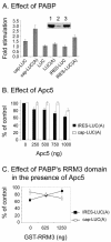
Effect of PABP and Apc5 on translation in vitro. (A) Krebs-2 cell-free translation reaction mixtures that had been pretreated with GST-Paip2 for PABP depletion or with GST as a control were programmed with 100 ng of the indicated transcripts at 37°C for 90 min followed by measurements of firefly luciferase activity. Immunoblot analysis of the translation extracts using antibodies specific for PABP is shown at the top: lane 1, untreated; lane 2, treated with GST-Paip2; lane 3, treated with GST. For each transcript, the value obtained in the GST-treated extract was divided by the value obtained in the PABP-depleted extract. The stimulation values represent the average ± standard error of three independent duplicate experiments. (B) Krebs-2 cell-free translation reaction mixtures preincubated for 30 min at room temperature with the indicated amounts of purified HIS-Apc5 were programmed with 50 ng of the indicated transcripts for a further 50-min incubation at 37°C. Firefly luciferase activity in the absence of Apc5 was set as 100%. The values represent the average ± standard error of three independent experiments. (C) Krebs-2 cell-free translation reaction mixtures preincubated for 30 min at room tem-perature with 1 μg of HIS-Apc5 and the indicated amounts of purified GST-RRM3 were programmed with 50 ng of the indicated transcripts for a further 50-min incubation at 37°C. Firefly luciferase activity in the absence of HIS-Apc5 and GST-RRM3 was set as 100%. The values represent the average ± standard error of three independent experiments.
FIG. 5.
FIG. 5.
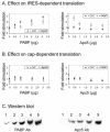
Apc5 represses IRES-mediated translation in a PABP-dependent manner. HEK293 cells were infected with vTF7-3 and cotransfected with 0.4 μg of pLPL, expressing PDGF2 IRES between Renilla and firefly luciferases (10), along with increasing amounts of pcDNA-hPABP or pcDNA-Apc5. The total amount of transfected plasmids was kept constant by compensation with pcDNA-CAT. As indicated, to each pcDNA-hPABP sample was added an additional 1 μg of either pcDNA-Apc5 (open squares) or pcDNA-CAT (solid circles) and to each pcDNA-Apc5 sample was added an additional 1 μg of either pcDNA-hPABP (open squares) or pcDNA-CAT (solid circles). Twenty-four hours after transfection, cells were harvested and assayed for the activity of Renilla (first cistron) (B) and firefly (second, IRES-controlled cistron) (A) luciferases. Basal activity of firefly luciferase, in the absence of additional Apc5 or PABP, was set as 1. The values represent the average ± standard error of three independent experiments. (C) Cells transfected with 0, 0.5, 1, 2, or 3 μg (lanes 1 to 5, respectively) of PABP- or Apc5-expressing plasmid were analyzed by immunoblotting with antibodies specific for PABP or Apc5 as indicated.
FIG. 6.
FIG. 6.
Apc5 is associated with other complexes, aside from APC/C. (A) Forty micrograms of proteins of the cytoplasmic S100 (C), RSW (R), and nuclear extracts (N) from control (−TPA) or differentiated (+TPA) K562 cells prepared as previously described (31) was immunoblotted with antibodies specific for Apc5, Apc3, or ribosomal protein S6 (rpS6). (B) Gel filtration chromatography. Total soluble proteins from logarithmically growing K562 cells lysed in the presence of a high (top panel) or low (bottom panel) concentration of detergents were separated by Superose-6 gel filtration chromatography. Equal volumes from each fraction were immunoblotted with antibodies specific for Apc5, Apc3, or rpS6. The elution point of the indicated molecular masses was calculated by using Unicorn software based on the elution points of thyroglobulin (669 kDa), apoferritin (443 kDa), catalase (232 kDa), and bovine serum albumin (66 kDa), which were used as molecular mass markers. (C) Density sucrose gradients. Twenty-five A260 units of K562 cells was resolved on a 5 to 45% sucrose gradient as described in Materials and Methods. Peaks containing the 40S, 60S, and 80S ribosomal subunits are indicated. Portions of the fractions were analyzed by immunoblotting with antibodies specific for Apc3, Apc5, or eIF3.
FIG. 7.
FIG. 7.
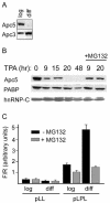
Apc5 is degraded upon megakaryocytic differentiation. (A) Apc5, but not Apc3, is degraded during differentiation. Fifty micrograms of total cell extract from logarithmically growing (log) or megakaryocytic differentiated (diff) K562 cells was separated by SDS-PAGE (10% polyacrylamide) and immunoblotted with antibodies specific for Apc5 or Apc3. (B) Proteasome inhibitor abolishes the differentiation-induced Apc5 degradation. K562 cells were treated with 5 nM TPA for 6 h followed by further incubation in medium containing 5 nM TPA with or without 30 μM MG132. At the indicated time points of TPA treatment (corresponding to 3 or 14 h of MG132 treatment), cells were harvested and 50 μg of total cell protein was immunoblotted with antibodies specific for Apc5, PABP, or hnRNP-C. (C) Proteasome inhibitor abolished the differentiation-induced IRES activation. K562 cells were transfected with pLPL expressing PDGF2 IRES between Renilla and firefly luciferases or the IRES-less vector pLL. Twenty-four hours after transfection, the cells were treated with 5 nM TPA, and 9 h later, MG132 was added to a final concentration of 30 μM. Forty-eight hours after transfection, cells were harvested and assayed for the activity of Renilla (R) (first cistron) and firefly (F) (second, IRES-controlled cistron) luciferases. The absolute values are presented in Table 1. The F/R values represent the firefly/Renilla activity ratio (arbitrary units) and are the average ± standard error of three independent experiments.
FIG. 8.
FIG. 8.
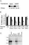
Apc5 overexpression inhibits differentiation-induced IRES activation. K562 cells were transfected with the IRES-less vector pLL or with pLPL, pLVL, pLML, pLXL, and pLEL harboring the IRESs of PDGF2, VEGF, c-myc, Xiap, and EMCV, respectively, between Renilla (R) and firefly (F) luciferase coding regions (10). Each of the bicistronic vectors was cotransfected along with pEGFP or pcDNA3-Apc5, expressing EGFP or Apc5, respectively. Twenty-four hours after transfection, cells were incubated under normal or differentiation conditions (5 nM TPA). (A) At the indicated time points along the differentiation process, the transfected cells were subjected to immunoblot analysis using anti-Apc5 antibodies. (B) Control and 48-h TPA-treated cells were harvested and assayed for the activity of Renilla (R) (first cistron) and firefly (F) (second, IRES-controlled cistron) luciferases. The D-IRES value is the F/R ratio in differentiated cells relative to that in control cells. The D-IRES value of each IRES in the presence EGFP was set as 1 (solid bars). The D-IRES values in the presence of Apc5 (open bars) represent the average ± standard error of three independent experiments. (C) Apc5 mRNA expression in different tissues. A multiple-tissue Northern blot (human 12-lane MTN #7780-1; Clontech) containing equal amounts of poly(A)+ from specific tissues (adjusted to β-actin hybridization signal) was hybridized with labeled Apc5 cDNA. RNA size markers (in kilobases) are indicated.

References

    1. Bentley, A. M., B. C. Williams, M. L. Goldberg, and A. J. Andres. 2002. Phenotypic characterization of Drosophila ida mutants: defining the role of APC5 in cell cycle progression. J. Cell Sci. 115:949-961. - PubMed
    1. Bergamini, G., T. Preiss, and M. W. Hentze. 2000. Picornavirus IRESes and the poly(A) tail jointly promote cap-independent translation in a mammalian cell-free system. RNA 6:1781-1790. - PMC - PubMed
    1. Bernstein, J., O. Sella, S. Y. Le, and O. Elroy-Stein. 1997. PDGF2/c-sis mRNA leader contains a differentiation-linked internal ribosomal entry site (D-IRES). J. Biol. Chem. 272:9356-9362. - PubMed
    1. Burd, C. G., E. L. Matunis, and G. Dreyfuss. 1991. The multiple RNA-binding domains of the mRNA poly(A)-binding protein have different RNA-binding activities. Mol. Cell. Biol. 11:3419-3424. - PMC - PubMed
    1. Cornelis, S., Y. Bruynooghe, G. Denecker, S. Van Huffel, S. Tinton, and R. Beyaert. 2000. Identification and characterization of a novel cell cycle-regulated internal ribosome entry site. Mol. Cell 5:597-605. - PubMed

Publication types

MeSH terms

Substances

LinkOut - more resources