. 2004 May;24(9):3633-47.
doi: 10.1128/MCB.24.9.3633-3647.2004.

Adapter protein SH2-B beta undergoes nucleocytoplasmic shuttling: implications for nerve growth factor induction of neuronal differentiation

Affiliations

Adapter protein SH2-B beta undergoes nucleocytoplasmic shuttling: implications for nerve growth factor induction of neuronal differentiation

Linyi Chen et al. Mol Cell Biol. 2004 May.

Abstract

The adapter protein SH2-B has been shown to bind to activated nerve growth factor (NGF) receptor TrkA and has been implicated in NGF-induced neuronal differentiation and the survival of sympathetic neurons. However, the mechanism by which SH2-B enhances and maintains neurite outgrowth is unclear. We examined the ability of truncation mutants to regulate neuronal differentiation and observed that certain truncation mutants localized in the nucleus rather than in the cytoplasm or at the plasma membrane as reported for wild-type SH2-B beta. Addition of the nuclear export inhibitor leptomycin B caused both overexpressed wild-type and endogenous SH2-B beta to accumulate in the nucleus of both PC12 cells and COS-7 cells as did deletion of a putative nuclear export sequence (amino acids 224 to 233) or mutation of two critical lysines in that sequence. Deleting or mutating the nuclear export signal caused SH2-B beta to lose its ability to enhance NGF-induced differentiation of PC12 cells. Neither the NGF-induced phosphorylation of ERKs 1 and 2 nor their subcellular distribution was altered in PC12 cells stably expressing the nuclear export-defective SH2-B beta(L231A, L233A). These data provide strong evidence that SH2-B beta shuttles constitutively between the nucleus and cytoplasm. However, SH2-B beta needs continuous access to the cytoplasm and/or plasma membrane to participate in NGF-induced neurite outgrowth. These data also suggest that the stimulatory effect of SH2-B beta on NGF-induced neurite outgrowth of PC12 cells is either downstream of ERKs or via some other pathway yet to be identified.


Figures

FIG. 1.
FIG. 1.
Effects of N-terminal truncation mutants of SH2-Bβ on neuronal differentiation. (A) Schematic representations of various N-terminal truncation mutants of SH2-Bβ fused to the C terminus of GFP. The proline-rich domain (P), pleckstrin homology domain (PH), and Src homology domain SH2 are depicted. The numbers are the amino acid numbers in the rat sequence of SH2-Bβ. (B) PC12 cells were transiently transfected with cDNAs encoding either GFP alone, full-length SH2-Bβ, or the SH2-Bβ mutants depicted in panel A and then treated with 100 ng of NGF per ml for 4 days. The percentage of differentiation was determined on day 4 as the number of transfected and differentiated cells divided by the number of transfected cells. A total of 150 to 200 transfected cells were counted for each condition in each experiment. The values are averages ± standard errors (error bars) from three different experiments.
FIG. 2.
FIG. 2.
Subcellular distribution of N-terminal truncation mutants of SH2-Bβ. (A) Schematic representations of various N-terminal truncation mutants of SH2-Bβ fused to the C terminus of GFP. The proline-rich domain (P), pleckstrin homology domain (PH), and Src homology domain SH2 are depicted. The numbers are the amino acid numbers in the rat sequence of SH2-Bβ. (B) PC12 cells were transiently transfected with cDNA encoding vector alone (G), full-length GFP-tagged SH2-Bβ (F), or a GFP-tagged mutant of SH2-Bβ shown in panel A, and designated by the number of its initial amino acid. Cells were lysed 18 h after transfection. Cell lysates were resolved by SDS-PAGE and immunoblotted with anti-SH2-B antibody. The migration positions of molecular weight (mw) standards (in thousands) are shown to the left of the gel. (C) PC12 cells were transiently transfected with cDNAs encoding GFP-tagged full-length SH2-Bβ [GFP-SH2-Bβ(1-670)] or an N-terminal truncation mutant of SH2-Bβ. The N-terminal truncation SH2-Bβ mutants GFP-SH2-Bβ(170-670), GFP-SH2-Bβ(200-670), GFP-SH2-Bβ(270-670), GFP-SH2-Bβ(397-670), and GFP-SH2-Bβ(504-670) were used. Cells were fixed 18 h after transfection with 4% paraformaldehyde, and the images were taken using epifluorescence microscopy. (D) PC12 cells were transiently transfected with cDNAs encoding GFP-tagged full-length SH2-Bβ or an N-terminal truncation mutant of SH2-Bβ and then treated with 100 ng of NGF per ml for 4 days. Images of live cells were taken on day 4 using epifluorescence microscopy.
FIG.3.
FIG.3.
Subcellular localization of the C-terminal truncation mutants of SH2-Bβ. (A) Schematic representations of various C-terminal truncation mutants of SH2-Bβ fused to the C terminus of GFP. (B) PC12 cells were transiently transfected with cDNAs encoding GFP-tagged full-length SH2-Bβ [GFP-SH2-Bβ(1-670)] or a C-terminal truncation mutant of SH2-Bβ shown in panel A. The C-terminal truncation SH2-Bβ mutants GFP-SH2-Bβ(1-631), GFP-SH2-Bβ(1-260), GFP-SH2-Bβ(1-200), and GFP-SH2-Bβ(1-150) were used. The cells were then treated with 100 ng of NGF per ml for 4 days. Images of live cells were taken on day 4. (C) COS-7 cells were transiently transfected with cDNAs encoding GFP-tagged full-length SH2-Bβ or a C-terminal truncation mutant of SH2-Bβ as indicated. Cells were fixed 18 h after transfection with 4% paraformaldehyde, and the images were taken using epifluorescence microscopy. DAPI images show nuclei.
FIG. 4.
FIG. 4.
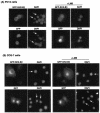
SH2-Bβ shuttles between the cytoplasm and nucleus. (A) PC12 cells were transiently transfected with cDNA encoding GFP-SH2-Bβ or GFP. Eighteen hours after transfection, cells were either mock treated (left panels) or treated with 20 nM leptomycin B (+LMB) for 3 h (right panels) before fixation. Images were then taken to determine the subcellular distribution of GFP-SH2-Bβ. DAPI images next to the fluorescence images showed the localization of the nucleus. (B) COS-7 cells were transiently transfected with GFP-SH2-Bβ or GFP. Eighteen hours after transfection, cells were either mock treated (left panels) or treated with 20 nM LMB for 5 h (right panels) before fixation. Images were then taken using epifluorescence microscopy. DAPI images showed the nucleus localization. (C) Quantification of GFP-SH2-Bβ distribution in the cytoplasm (C) or in the cytoplasm and nucleus (C+N) in either PC12 cells or COS-7 cells without or with LMB (+ LMB) treatment. A total of 17 to 43 cells were counted from two experiments per condition. (D) PC12 cells were incubated without or with LMB for 3h before fixation. Endogenous SH2-Bβ was detected by incubating cells with goat anti-SH2-Bβ antibody and then with rabbit anti-goat Alexa 488. The images were visualized using epifluorescence microscopy. The corresponding DAPI images show the nuclei. (E) COS-7 cells were incubated without or with LMB for 28 h before fixation. Endogenous SH2-Bβ was detected and visualized as described for panel D.
FIG. 4.
FIG. 4.
SH2-Bβ shuttles between the cytoplasm and nucleus. (A) PC12 cells were transiently transfected with cDNA encoding GFP-SH2-Bβ or GFP. Eighteen hours after transfection, cells were either mock treated (left panels) or treated with 20 nM leptomycin B (+LMB) for 3 h (right panels) before fixation. Images were then taken to determine the subcellular distribution of GFP-SH2-Bβ. DAPI images next to the fluorescence images showed the localization of the nucleus. (B) COS-7 cells were transiently transfected with GFP-SH2-Bβ or GFP. Eighteen hours after transfection, cells were either mock treated (left panels) or treated with 20 nM LMB for 5 h (right panels) before fixation. Images were then taken using epifluorescence microscopy. DAPI images showed the nucleus localization. (C) Quantification of GFP-SH2-Bβ distribution in the cytoplasm (C) or in the cytoplasm and nucleus (C+N) in either PC12 cells or COS-7 cells without or with LMB (+ LMB) treatment. A total of 17 to 43 cells were counted from two experiments per condition. (D) PC12 cells were incubated without or with LMB for 3h before fixation. Endogenous SH2-Bβ was detected by incubating cells with goat anti-SH2-Bβ antibody and then with rabbit anti-goat Alexa 488. The images were visualized using epifluorescence microscopy. The corresponding DAPI images show the nuclei. (E) COS-7 cells were incubated without or with LMB for 28 h before fixation. Endogenous SH2-Bβ was detected and visualized as described for panel D.
FIG. 5.
FIG. 5.
Subcellular distribution of GFP-SH2-Bβ(Δ198-268). (A) Schematic representation of the internal deletion mutant of SH2-Bβ fused to the C terminus of GFP, GFP-SH2-Bβ(Δ198-268). (B) PC12 cells were transiently transfected with cDNAs encoding either GFP-SH2-Bβ or GFP-SH2-Bβ(Δ198-268). Cells were fixed, and images were taken using epifluorescence microscopy. DAPI images next to the fluorescence images showed the nucleus localization. (C) PC12 cells were transiently transfected with cDNA encoding either GFP-SH2-Bβ or GFP-SH2-Bβ(Δ198-268) and then treated with 100 ng of NGF per ml for 4 days. Live images of differentiated PC12 cells were taken on day 4 to show the localization of GFP-SH2-Bβ or GFP-SH2-Bβ(Δ198-268). (D) COS-7 cells were transiently transfected with cDNA encoding either GFP-SH2-Bβ or GFP-SH2-Bβ(Δ198-268). Cells were fixed 18 h after transfection, and images were taken using epifluorescence microscopy. DAPI images next to the fluorescence images were used to show the nucleus localization.
FIG. 6.
FIG. 6.
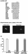
Point mutations in the putative NES motif in SH2-Bβ inhibit the nuclear export of SH2-Bβ. (A) Known NESs in various classes of proteins and the putative NES in SH2-B. The arrows point to the two leucines in SH2-Bβ being mutated. (B) PC12 cells were transiently transfected with the cDNA encoding the GFP-fused SH2-Bβ point mutant, SH2-Bβ(L231A, L233A). Cells were fixed 18 h after transfection (undifferentiated). Another set of cells was treated with 100 ng of NGF per ml for 4 days, and live images of the differentiated PC12 cells were taken. (C) Quantification of the localization of GFP-SH2-Bβ, GFP-SH2-Bβ(Δ198-268), or SH2-Bβ(L231A, L233A) in either the cytoplasm (C) or in both the cytoplasm and nucleus (C+N). A total of 61 to 173 differentiated PC12 cells from three experiments were counted for each construct.
FIG. 7.
FIG. 7.
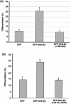
Effect of SH2-Bβ(Δ198-268) and SH2-Bβ(L231A, L233A) on neuronal differentiation. (A) PC12 cells were transiently transfected with the cDNA encoding the vector alone (GFP), GFP-SH2-Bβ, or GFP-SH2-Bβ(Δ198-268) as indicated. Cells were then treated with 100 ng of NGF per ml for 4 days, and the percentage of differentiation was determined as described in the legend to Fig. 1. A total of 300 to 450 transfected cells were counted for each condition in each experiment, and the values are averages ± standard errors (error bars) from three different experiments. (B) PC12 cells were transiently transfected with the cDNA encoding the vector alone, GFP-SH2-Bβ, or GFP-SH2-Bβ(L231A, L233A) as indicated. Cells were then treated with 100 ng of NGF per ml for 4 days, and the percentage of differentiation was determined as described in the legend to Fig. 1. A total of 333 to 870 transfected cells were counted for each condition in each experiment, and the values are averages ± standard deviations (error bars) from two different experiments.
FIG.8.
FIG.8.
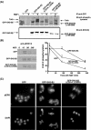
Effect of a nuclear export-defective mutant of SH2-Bβ on NGF signaling. (A) PC12 cells stably expressing either GFP, GFP-SH2-Bβ, GFP-SH2-Bβ(L231A, L233A), or GFP-SH2-Bβ(Δ198-268) as indicated were incubated in serum-free medium overnight and then pretreated with sodium vanadate for 1 h before stimulation without (−) with (+) 100 ng of NGF per ml for 10 min. Cell lysates were incubated with anti-GFP antibody and then with protein A agarose. The immunoprecipitated complex was resolved by SDS-PAGE and immunoblotted with antiphosphotyrosine antibody (top gel) and then reprobed with anti-SH2-B antibody (bottom gel). IP, immunoprecipitation; IB, immunoblotting. (B) PC12 cells stably expressing either GFP, GFP-SH2-Bβ, or GFP-SH2-Bβ(L231A, L233A) were incubated in serum-free medium overnight and stimulated with 100 ng of NGF per ml for 0, 10, 90, and 240 min (240′). Lysates from these cell lines expressing GFP, GFP-SH2-Bβ, or GFP-SH2-Bβ(L231A, L233A) were resolved by SDS-PAGE, and the blot was incubated with anti-phospho-ERKs 1 and 2 (anti-pERK1/2). The highest level of phosphorylated ERKs 1 and 2 in each experiment was set at 100. The values are the averages ± standard errors (error bars) from three different experiments. (C) PC12 stable cell lines were treated with 100 ng of NGF per ml for 1 h as described for panel B and then fixed and incubated with a rabbit antibody against active phosphorylated ERK (pERK) and then with goat anti-rabbit Alexa 555 antibody. DAPI images reveal the nuclei. The images were taken using epifluorescence microscopy.

Similar articles

Cited by

References

    1. Bardwell, A. J., L. J. Flatauer, K. Matsukuma, J. Thorner, and L. Bardwell. 2001. A conserved docking site in MEKs mediates high-affinity binding to MAP kinases and cooperates with a scaffold protein to enhance signal transmission. J. Biol. Chem. 276:10374-10386. - PMC - PubMed
    1. Bogerd, H. P., R. A. Fridell, R. E. Benson, J. Hua, and B. R. Cullen. 1996. Protein sequence requirements for function of the human T-cell leukemia virus type 1 Rex nuclear export signal delineated by a novel in vivo randomization-selection assay. Mol. Cell. Biol. 16:4207-4214. - PMC - PubMed
    1. Brunet, A., F. Kanai, J. Stehn, J. Xu, D. Sarbassova, J. V. Frangioni, S. N. Dalal, J. A. DeCaprio, M. E. Greenberg, and M. B. Yaffe. 2002. 14-3-3 transits to the nucleus and participates in dynamic nucleocytoplasmic transport. J. Cell Biol. 156:817-828. - PMC - PubMed
    1. Cheng, S., M. S. Geddis, and V. Rehder. 2002. Local calcium changes regulate the length of growth cone filopodia. J. Neurobiol. 50:263-275. - PubMed
    1. Choi, D. Y., J. J. Toledo-Aral, R. Segal, and S. Halegoua. 2001. Sustained signaling by phospholipase C-γ mediates nerve growth factor-triggered gene expression. Mol. Cell. Biol. 21:2695-2705. - PMC - PubMed

Publication types

MeSH terms

LinkOut - more resources