. 2004 May;24(9):3682-91.
doi: 10.1128/MCB.24.9.3682-3691.2004.

Epidermal growth factor receptor stimulation activates the RNA binding protein CUG-BP1 and increases expression of C/EBPbeta-LIP in mammary epithelial cells

Affiliations

Epidermal growth factor receptor stimulation activates the RNA binding protein CUG-BP1 and increases expression of C/EBPbeta-LIP in mammary epithelial cells

Brenda R Baldwin et al. Mol Cell Biol. 2004 May.

Abstract

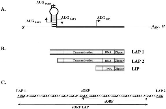
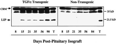
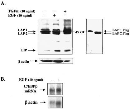
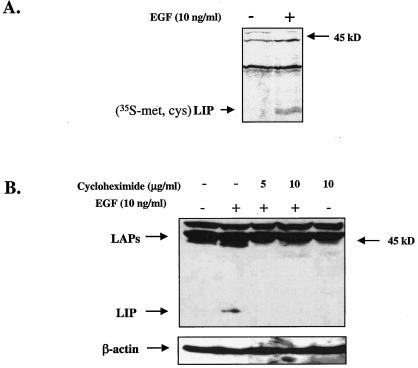
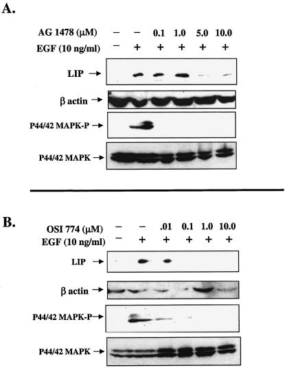
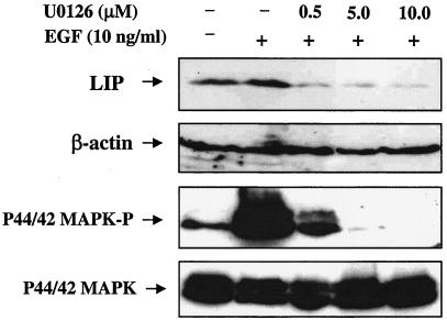
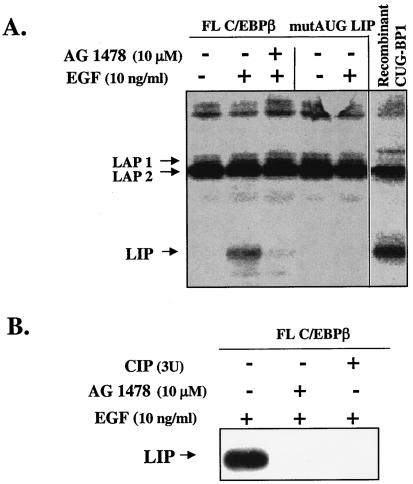
The transcription factor CCAAT/enhancer binding protein beta (C/EBP beta) is a key regulator of growth and differentiation in many tissues. C/EBP beta is expressed as several distinct protein isoforms (LAP1, LAP2, and LIP) whose expression is regulated by alternative translational initiation at downstream AUG start sites. The dominant-negative LIP isoform is predominantly expressed during proliferative cellular responses and is associated with aggressive tumors. In this study, we investigated a mechanism by which the LIP isoform is translationally regulated in mammary epithelial cells. We have demonstrated that LIP expression is increased in response to activation of the epidermal growth factor receptor (EGFR) signaling pathway and that the increased expression of LIP is regulated in part by an RNA binding protein referred to as CUG repeat binding protein (CUG-BP1). Our data demonstrate that EGFR signaling results in the phosphorylation of CUG-BP1 and this leads to an increase in the binding of CUG-BP1 to C/EBP beta mRNA and elevated expression of the LIP isoform. Phosphorylation is necessary for the binding activity of CUG-BP1 and the consequent increase in LIP expression, as determined by binding assays and a cell free, transcription-coupled translation system. CUG-BP1 is thus a previously unidentified downstream target of EGFR signaling and represents a new translational regulator of LIP expression in human mammary epithelial cells.


Figures

FIG. 1.
FIG. 1.
Schematic of C/EBPβ mRNA and protein isoforms. (A) Line drawing of C/EBPβ mRNA. To show the hairpin structure and translation start sites in greater detail, the diagram has not been drawn to scale. The thin line represents the untranslated noncoding sequence, and the thick line represents the translated coding sequence. (B) Schematic of the predominant C/EBPβ protein isoforms. The transactivation, DNA binding (DNA), and leucine zipper (zipper) regions are indicated. (C) Ribonucleotide sequence of the murine C/EBPβ hairpin region from the LAP1 AUG codon to the LAP2 AUG codon to show the sequences spanned by the murine oligomers used in the RNA binding assay. The mouse and human ribonucleotide sequences are 81% identical in this region.
FIG. 2.
FIG. 2.
LIP expression is increased in WAP-TGF-α mice after transgene stimulation. Western blot analysis of whole-cell extracts (100 μg) prepared from mammary glands removed (8 to 90 days) after implantation of pituitary isografts into the thoracic glands of TGF-α transgenic mice (left panel) and nontransgenic siblings (right panel). The days post pituitary isografting are indicated. Whole-cell extracts from a non-pituitary-isografted TGF-α tumor (T) serves as a positive control for LIP overexpression. The blots were overexposed in an effort to detect LIP in the nontransgenic blot. Cross-reactive material (CRM) serves as an estimate for the total protein loaded and transferred per well.
FIG. 3.
FIG. 3.
Protein expression of LIP is induced in MCF10A cells after treatment with EGF or TGF-α, but transcriptional regulation of C/EBPβ is relatively unaffected. (A, left side) Western blot analysis of LIP in MCF10A cells treated with EGF or TGF-α. Cells were serum starved for 24 h and then treated with human EGF (10 ng/ml) or TGF-α (10 ng/ml). Cells were harvested 16 h after addition of ligand, and 100 μg of protein from whole-cell extracts was subjected to SDS-PAGE and Western blot analysis. Rehybridization of the blot with a β-actin polyclonal antibody served as an estimate of the total amount of protein loaded and transferred per well. (A, right side) Western blot analysis of 293 cells transfected with LAP1 or LAP2 constructs that were Flag tagged at the amino terminus. LAP isoforms were immunodetected with Flag antibody (Sigma) and serve as controls to aid in the identification of the endogenous LAP isoforms that often migrate with cross-reactive material and are often difficult to resolve on Western blots. (B) Northern blot analysis of poly(A)+ mRNA (7 μg) isolated from EGF-treated and nontreated MCF10A cells hybridized with a cDNA probe to human C/EBPβ. Blots were stripped and rehybridized with a cDNA for β-actin to estimate RNA loading per well.
FIG. 4.
FIG. 4.
EGF-induced expression of LIP is the result of nascent protein synthesis. (A) MCF10A cells were serum starved for 24 h and then pulsed with [35S]Met-Cys for 3.5 h (10 ng/ml). Cells were chased with unlabeled Met-Cys medium and then harvested 24 h after addition of EGF. 35S-labeled proteins were immunoprecipitated with a C/EBPβ polyclonal antibody and analyzed via SDS-PAGE and autoradiography. (B) Western blot analysis of whole-cell protein extracts (100 μg) from MCF10A cells that were treated with cycloheximide (5 to 10 μg/ml) for 30 min prior to the 16-h EGF treatment (10 μg/ml). The blot was stripped and reprobed with a β-actin polyclonal antibody to estimate loading and transfer of proteins (bottom).
FIG. 5.
FIG. 5.
Expression of LIP is decreased in MCF10A cells after treatment with EGFR antagonists. (A) MCF10A cells were serum starved for 24 h and then treated with increasing concentrations of AG1478 for 30 min prior to addition of EGF (10 ng/ml). Cells were then harvested at either 5 min (for p44/42 MAPK detection; lower panels) or 16 h (for LIP detection; upper panel) after EGF stimulation. Whole-cell extracts (100 μg) were analyzed via SDS-PAGE and Western blotting. C/EBPβ blots were stripped and reprobed with a polyclonal antibody to β-actin (middle panel) to estimate loading and transfer of proteins. (B) Dose response of MCF10A cells to treatment with OSI-774. Experiment was conducted as described for panel A.
FIG. 6.
FIG. 6.
Effect of inhibition of p44/42 MAPK signaling on the EGF-induced expression of LIP. Inhibition of p44/42 MAPK activation with U0126 reduces expression of LIP. MCF10A cells were serum starved for 24 h and then treated with increasing concentrations of U0126, a potent MEK1/2 inhibitor, for 30 min prior to addition of EGF (10 ng/ml). Cells were then harvested at either 5 min (for p44/42 MAPK-P and total p44/42 MAPK detection; lower panels) or 16 h (for LIP detection; upper panel) after addition of EGF. Whole-cell extracts (100 μg) were analyzed via SDS-PAGE and Western blotting. The C/EBPβ blot was stripped and incubated with a polyclonal antibody to β-actin to estimate loading and transfer of proteins.
FIG. 7.
FIG. 7.
EGFR signaling regulates the phosphorylation status of CUG-BP1. (A and B) Western blot analysis of whole-cell protein extracts demonstrates that treatment with EGF leads to the appearance of a CUG-BP1 doublet (A and B, lane 2) that is reduced to a single band by treatment with either OSI-774 (A, lanes 4 and 5) or CIP (B, lanes 3 and 4). (C) EGF stimulation leads to increased incorporation of 32P into CUG-BP1. EGF-treated MCF10A cells were metabolically labeled with 32Pi, and CUG-BP1 was immunoprecipitated and analyzed by PAGE and autoradiography. A basal level of phosphorylation of CUG-BP1 is observed in the nontreated cells (−), which is increased upon stimulation with EGF (+).
FIG. 8.
FIG. 8.
The binding activity of CUG-BP1 is regulated by EGFR-induced phosphorylation. (A and B) Binding of CUG-BP1 to C/EBPβ mRNA is increased by stimulation of cells with EGF and inhibited by the EGF antagonists AG1478 and OSI-774 and the MEK1/2 inhibitor U0126. MCF10A cells were serum starved for 24 h and treated with either AG1478 (A), OSI-774 (B), or U0126 (C, left part) for 30 min prior to addition of EGF. Cells were harvested at 16 h, and whole-cell protein extracts were incubated with [γ-32P]ATP-labeled C/EBPβ RNA probes (Fig. 1C) and cross-linked with UV light. RNA-bound proteins were analyzed via electrophoresis on 8 to 16% denaturing polyacrylamide gradient gels, transferred onto nitrocellulose membranes, and visualized by autoradiography. (C, right part) CUG-BP1 interacts specifically with GCN repeats located in the 5′ region of C/EBPβ mRNA. Addition of an excess of an unlabeled RNA oligonucleotide (sORF), but not that of an RNA oligonucleotide (AU-rich), abolished the binding of CUG-BP1 to the labeled (sORF-LAP) probe, as detected by PAGE and autoradiography. (D) Phosphatase treatment of CUG-BP1 abolishes binding to C/EBPβ mRNA, as determined by UV cross-link-immunoprecipitation assay. As described above, MCF10A cells were stimulated with EGF and whole-cell extracts were treated with phosphatase. CUG-BP1 was then immunoprecipitated from both EGF and non-EGF-stimulated cells (lanes 1 and 2) and from CIP-treated, EGF-activated extracts (lanes 3 and 4). The immunoprecipitate was incubated with a [γ-32P]ATP-labeled C/EBPβ sORF probe and analyzed via SDS-PAGE and autoradiography.
FIG. 9.
FIG. 9.
CUG-BP1 immunoprecipitated from EGF-treated MCF10A cells induces the translation of LIP in a cell-free system. (A) CUG-BP1 was immunoprecipitated from whole-cell extracts of MCF10A cells that were treated for 16 h with EGF, EGF plus AG1478, or vehicle only. The immunoprecipitated CUG-BP1 protein was added to the rabbit RL in the presence of [35S]methionine and either a full-length WT C/EBPβ construct (FL) or a mutated construct containing an ATG-to-TTG mutation at the LIP AUG codon (mutAUG). The lane designated recombinant CUG-BP1 (see Materials and Methods) is a positive control containing bacterially expressed CUG-BP1 that has been activated with HeLa cell extracts, immunoprecipitated, and added to the 35S-labeled RL programmed with the FL C/EBPβ construct. After translation, the C/EBPβ isoforms were immunoprecipitated, separated by SDS-PAGE, and visualized by autoradiography. (B) CUG-BP1 was immunoprecipitated from whole-cell extracts as described in panel A. The immunoprecipitate from the EGF-treated cells was treated with CIP, washed, and transferred to the TnT reaction mixture, which contained a full-length, WT C/EBPβ cDNA construct. Translated proteins were subjected to Western blot analysis with a polyclonal antibody to C/EBPβ.

Similar articles

Cited by

References

    1. Alaoui-Jamali, M. A., D. J. Song, N. Benlimame, L. Yen, X. Deng, M. Hernandez-Perez, and T. Wang. 2003. Regulation of multiple tumor microenvironment markers by overexpression of single or paired combinations of ErbB receptors. Cancer Res. 63:3764-3774. - PubMed
    1. Badley, J. E., G. A. Bishop, T. St John, and J. A. Frelinger. 1988. A simple, rapid method for the purification of poly A+ RNA. BioTechniques 6:114-116. - PubMed
    1. Baer, M., and P. F. Johnson. 2000. Generation of truncated C/EBPβ isoforms by in vitro proteolysis. J. Biol. Chem. 275:26582-26590. - PubMed
    1. Bundy, L. M., and L. Sealy. 2003. CCAAT/enhancer binding protein beta (C/EBPβ)-2 transforms normal mammary epithelial cells and induces epithelial to mesenchymal transition in culture. Oncogene 22:869-883. - PubMed
    1. Calkhoven, C. F., C. Muller, and A. Leutz. 2000. Translational control of C/EBPα and C/EBPβ isoform expression. Genes Dev. 14:1920-1932. - PMC - PubMed

Publication types

MeSH terms