. 2004 May;24(9):3712-9.
doi: 10.1128/MCB.24.9.3712-3719.2004.

Identification of the XPG region that causes the onset of Cockayne syndrome by using Xpg mutant mice generated by the cDNA-mediated knock-in method

Affiliations

Identification of the XPG region that causes the onset of Cockayne syndrome by using Xpg mutant mice generated by the cDNA-mediated knock-in method

Naoko Shiomi et al. Mol Cell Biol. 2004 May.

Abstract

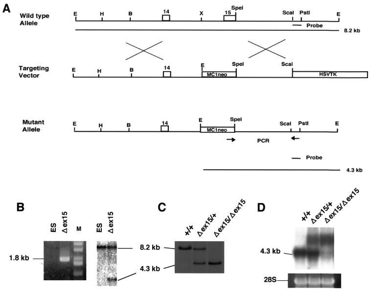
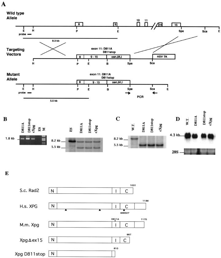
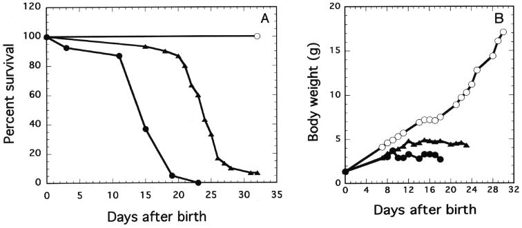
In addition to xeroderma pigmentosum (XP), mutations in the human XPG gene cause early onset of Cockayne syndrome (CS) in some patients (XPG/CS). The CS-causing mutations in such patients all produce truncated XPG proteins. To test the hypothesis that the CS phenotype, with characteristics such as growth retardation and a short life span in XPG/CS patients, results from C-terminal truncations, we constructed mutants with C-terminal truncations in mouse XPG (Xpg) (from residue D811 to the stop codon [XpgD811stop] and deletion of exon 15 [Xpg Delta ex15]). In the XpgD811stop and Xpg Delta ex15 mutations, the last 360 and 183 amino acids of the protein were deleted, respectively. To generate Xpg mutant mice, we devised the shortcut knock-in method by replacing genomic DNA with a mutated cDNA fragment (cDNA-mediated knock in). The control mice, in which one-half of Xpg genomic DNA fragment was replaced with a normal Xpg cDNA fragment, had a normal growth rate, a normal life span, normal sensitivity to UV light, and normal DNA repair ability, indicating that the Xpg gene partially replaced with the normal cDNA fragment retained normal functions. The XpgD811stop homozygous mice exhibited growth retardation and a short life span, but the Xpg Delta ex15 homozygous mice did not, indicating that deletion of the last 360 amino acids results in the CS phenotype but deletion of the last 183 amino acids does not. The XpgD811stop homozygous mice, however, exhibited a slightly milder CS phenotype than did the Xpg null mutant mice, indicating that the XpgD811stop protein still retains some Xpg function that affects the severity of the CS phenotype.


Figures

FIG. 1.
FIG. 1.
Generation of Xpg-deficient (Δex15) mice. (A) Schematic of the deletion mutation at the Xpg locus. Exons 14 and 15 of the Xpg gene are represented as boxes. PCR primers are indicated by arrows. The 3′ external probe used for Southern blot analysis is indicated by a solid bar corresponding to the ScaI-PstI fragment on the wild-type map at the top of panel A, and the diagnostic fragments of 4.3 and 8.2 kb are indicated by solid lines at the bottom of panel A. Abbreviations: B, BamHI; E, EcoRI; H, HindIII; X, XhoI. (B) PCR and Southern blot analyses of the targeted ES clone, G138. The size of the predicted PCR product for the mutant allele was 1.8 kb, and no PCR product was amplified from the wild-type allele. Southern blot analysis with the 3′ external probe also detected the predicted restriction fragments shown in panel A. Lanes: M, size markers; ES, ES cells used as a control. (C) Southern blot analyses of offspring from intercrosses between chimeric males and C57BL/6J females. Lanes: +/+, wild type; +/Δex15, heterozygote; Δex15/Δex15, homozygous XpgΔex15 mutant. (D) Northern blot analysis of total RNA from newborn mice derived from a heterozygous intercross, with Xpg cDNA as the probe. As a loading control, the 28S rRNA band is shown.
FIG. 2.
FIG. 2.
Generation of a truncation or base substitution mutation in the Xpg gene by the cDNA-mediated knock-in method. (A) Schematic of generation of mutations at the Xpg locus by the cDNA-mediated knock-in method. Exons 8 to 15 are shown. PCR primers are indicated by arrows. The 5′ probe used for Southern blot analysis is indicated by a solid bar on the wild-type map at the top of panel A, and the diagnostic fragments of 5.5 and 8.3 kb are indicated by solid lines at the bottom of panel A. Abbreviations: B, BamHI; E, EcoRI; H, HindIII; P, PstI. (B) PCR and Southern blot analyses of the targeted ES clones, ES-D811A, ES-D811stop, and ES-cXpg. The size of the predicted PCR product for mutant alleles was 1.8 kb, and no PCR product was amplified from the wild-type allele. Southern blot analysis with the 5′ probe also detected the predicted restriction fragments shown in panel A. Lanes: M, size markers; ES, ES cells used as a control. (C) Southern blot analyses of offspring from intercrosses between chimeric males and C57BL/6J females. +/+ represents the wild type (W.T.). cXpg/cXpg, D811A/D811A, and D811stop/D811stop represent homozygous cXpg, XpgD811A, and XpgD811stop mutants, respectively. (D) Northern blot analysis of total RNA from newborn mice, which were derived from heterozygous intercrosses and were determined to be homozygous mutants, with Xpg cDNA as the probe. As a loading control, the 28S rRNA band is shown. (E) Schematic of mutations in the Xpg protein. Arrows indicate the locations of mutations in XPG where the protein terminates in three XPG/CS patients. N and I indicate the highly conserved N-terminal and internal regions. C indicates the moderately conserved C-terminal region. The Xpg and S. cerevisiae (S.c.) Rad2 proteins have 45.5, 52.6, and 34.2% identical residues in the N, I, and C regions, respectively. In the XpgΔex15 and XpgD811stop mutant proteins, the last 183 and 360 amino acids were deleted, respectively. In the XpgD811A mutation, the highly conserved D811 residue of the Xpg protein was replaced with alanine. H.s., Homo sapiens; M.m., Mus musculus.
FIG. 3.
FIG. 3.
Survival curves of embryonic fibroblasts derived from Xpg mutant mice. Each point represents an average of three independent experiments with wild-type (○), cXpg (□), XpgD811A (▪), XpgD811stop (▴), XpgΔex15 (▵), or XpgΔ (•) homozygous cells. Error bars show the standard deviation derived from at least three independent experiments.
FIG. 4.
FIG. 4.
Kinetics of UV light-induced DNA damage removal in embryonic fibroblasts derived from wild-type (○), cXpg (□), XpgD811A (▪), XpgD811stop (▴), XpgΔex15 (▵), or XpgΔ (•) homozygous mice. The kinetics of 6-4 photoproduct (6-4PP) (A) and cyclobutane pyrimidine dimer (CPD) (B) removal are presented. Each point is the average of triplicate wells. Error bars show the standard deviation derived from at least three independent experiments.
FIG. 5.
FIG. 5.
Growth rate and life span of Xpg mutant mice. Survival curves (A) and average body weights (B) of wild-type (○), XpgD811stop (▴), and XpgΔ (•) homozygous mice are shown. Since the survival curves and body weights of cXpg, XpgD811A, and XpgΔex15 homozygous mice are similar to those of wild-type mice, the plots for these mice were omitted.

References

    1. Aboussekhra, A., and R. D. Wood. 1994. Repair of UV-damaged DNA by mammalian cells and Saccharomyces cerevisiae. Curr. Opin. Genet. Dev. 4:212-220. - PubMed
    1. Aboussekhra, A., M. Biggerstaff, M. K. Shivji, J. A. Vilpo, V. Monocollin, V. N. Podust, M. Protic, U. Hubscher, J. M. Egly, and R. D. Wood. 1995. Mammalian DNA nucleotide excision repair reconstituted with purified protein components. Cell 80:859-868. - PubMed
    1. Bambara, R. A., R. S. Murante, and L. A. Henricksen. 1997. Enzymes and reactions at the eukaryotic DNA replication fork. J. Biol. Chem. 272:4647-4650. - PubMed
    1. Bootsma, D., and J. H. J. Hoeijmakers. 1993. Engagement with transcription. Nature 363:114-115. - PubMed
    1. Bradsher, J., J. Auriol, L. P. de Santis, S. Iban, J. L. Vonesh, I. Grummt, and J. M. Egly. 2002. CSB is a component of RNA pol I transcription. Mol. Cell 10:819-829. - PubMed

Publication types

MeSH terms