. 2004 May;24(9):3757-68.
doi: 10.1128/MCB.24.9.3757-3768.2004.

HRC is a direct transcriptional target of MEF2 during cardiac, skeletal, and arterial smooth muscle development in vivo

Affiliations

HRC is a direct transcriptional target of MEF2 during cardiac, skeletal, and arterial smooth muscle development in vivo

Joshua P Anderson et al. Mol Cell Biol. 2004 May.

Abstract

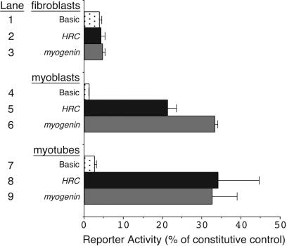
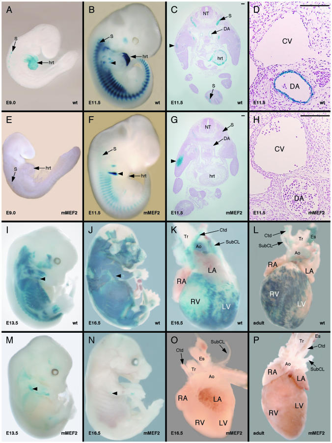
The HRC gene encodes the histidine-rich calcium-binding protein, which is found in the lumen of the junctional sarcoplasmic reticulum (SR) of cardiac and skeletal muscle and within calciosomes of arterial smooth muscle. The expression of HRC in cardiac, skeletal, and smooth muscle raises the possibility of a common transcriptional mechanism governing its expression in all three muscle cell types. In this study, we identified a transcriptional enhancer from the HRC gene that is sufficient to direct the expression of lacZ in the expression pattern of endogenous HRC in transgenic mice. The HRC enhancer contains a small, highly conserved sequence that is required for expression in all three muscle lineages. Within this conserved region is a consensus site for myocyte enhancer factor 2 (MEF2) proteins that we show is bound efficiently by MEF2 and is required for transgene expression in all three muscle lineages in vivo. Furthermore, the entire HRC enhancer sequence lacks any discernible CArG motifs, the binding site for serum response factor (SRF), and we show that the enhancer is not activated by SRF. Thus, these studies identify the HRC enhancer as the first MEF2-dependent, CArG-independent transcriptional target in smooth muscle and represent the first analysis of the transcriptional regulation of an SR gene in vivo.


Figures

FIG. 1.
FIG. 1.
An evolutionarily conserved noncoding sequence resides in the upstream region of the HRC gene. (A) Schematic representation of the human HRC upstream region. The HRC upstream region (−2609 to +117) was cloned as a BamHI-Psp1406I fragment into CAT and β-galactosidase reporter plasmids such that transcription would start from the HRC transcriptional start site (red arrow) and translation would initiate in the reporter cDNA. Green box, evolutionarily conserved region; black arrow, HRC translational start site; B, BamHI; N, NcoI; Bg, BglII; P, Psp1406I. (B) Sequence alignment of the evolutionarily conserved element from the HRC upstream region from −608 to −468, relative to the transcriptional start site, in the human sequence. The box denotes the evolutionarily conserved MEF2 site.
FIG. 2.
FIG. 2.
The HRC upstream region contains promoter and enhancer sequences sufficient to direct muscle-specific expression. HRC-CAT (lanes 2, 5, and 8), myogenin-CAT (lanes 3, 6, and 9), and the promoterless CAT-Basic (lanes 1, 4, and 7) reporter plasmids were transfected into fibroblasts (lanes 1 to 3), myoblasts (lanes 4 to 6), or myotubes (lanes 7 to 9). The HRC and myogenin reporter plasmids exhibited no significant activity over background in nonmuscle fibroblasts (lanes 2 and 3), but both reporters were robustly active in myoblasts (lanes 5 and 6) and myotubes (lanes 8 and 9). Data are expressed as a percentage of the activity obtained using a constitutively active SV40-CAT plasmid in each cell type. The data shown represent the mean values obtained in five independent transfections and analyses. Error bars represent the standard errors of the means.
FIG. 3.
FIG. 3.
The HRC enhancer directs cardiac, skeletal, and arterial smooth muscle expression in transgenic mouse embryos. A 2,726-bp fragment of the human HRC gene was fused to a lacZ reporter plasmid and used to generate transgenic mice. This fragment of the HRC gene was sufficient to direct expression to all three muscle lineages in the mouse embryo in the pattern of endogenous HRC. (A to D) Representative X-Gal-stained transgenic embryos are shown at 8.5 dpc (A), 9.5 dpc (B), 11.5 dpc (C), and 13.5 dpc (D). The embryos in panels C and D have been cleared in a 1:1 mixture of benzyl alcohol and benzyl benzoate to help visualization of internal structures. Expression was evident at 8.5 dpc in the heart (hrt) and in the myotomal compartment of the somites (S) and by 11.5 dpc in arterial smooth muscle. No expression was observed in venous smooth muscle or in other smooth muscle cell types. (E to G) Transverse sections from X-Gal-stained transgenic embryos at 11.5 dpc. Expression was evident in somites, heart, and arterial vascular smooth muscle, including the dorsal aorta (DA) and branchial arch arteries (BAA). By contrast, expression was not observed in venous smooth muscle, including the cardinal veins (CV). No expression was observed in the smooth muscle of the trachea (Tr) or esophagus (Es) (G). Expression in the heart was restricted to the ventricles (E and F). Bar, 100 μm. (H) The heart and associated vasculature removed from an HRC-lacZ transgenic embryo at 13.5 dpc and stained with X-Gal. Expression in the heart at 13.5 dpc was restricted to the ventricles. Expression was also evident in arterial vascular smooth muscle, including the aorta (Ao) and subclavian (SubCL) and carotid arteries (Ctd). LA, left atrium; LV, left ventricle; NT, neural tube; RA, right atrium; RV, right ventricle. Seven independent transgenic lines all displayed nearly identical patterns of expression.
FIG. 4.
FIG. 4.
Deletional analysis of the HRC upstream region identified a conserved region required for expression in vivo. Schematic representations of the various deletion constructs of the HRC upstream region analyzed in these studies are shown in the center. Red arrow, HRC transcriptional start site; green arrow, transcriptional start site directed by the heterologous HSP68 promoter; blue arrow, lacZ translational start site; B, BamHI; N, NcoI; Bg, BglII; P, Psp1406I. Construct number and nucleotides, relative to the HRC transcriptional start site at +1, are indicated on the left. Expression of lacZ in somites, limbs, heart, and arteries is noted in the columns to the right. +++, very robust, easily detectable expression; +, very weak expression; −, a complete lack of detectable expression. The column on the far right indicates the number of independent transgenic lines or F0 transgenic embryos that expressed lacZ in the indicated pattern as a fraction of the total number of transgene-positive F0 embryos or lines examined. For HSP68/510-770 (construct 5), three of the five lines examined expressed lacZ in the indicated pattern. The other two F0 transgenic embryos showed no expression of lacZ.
FIG. 5.
FIG. 5.
The conserved region of the HRC enhancer contains a high-affinity, functional MEF2 site. (A) MEF2 binds specifically to the HRC MEF2 site in vitro. MEF2A was transcribed and translated in vitro and used in EMSA analyses with radiolabeled double-stranded oligonucleotides representing the HRC MEF2 site (lanes 1 to 6) or a mutant version of the HRC MEF2 site (lanes 7 and 8). MEF2 efficiently bound to the HRC MEF2 site (lane 2) but failed to bind to the mutant MEF2 site (lane 8). Binding of MEF2 to the HRC MEF2 site was specific, since a 100-fold excess of unlabeled HRC MEF2 site efficiently competed for binding (lane 3) but a mutant version of the HRC MEF2 site (mHRC) failed to compete for binding even at a 100-fold excess (lane 4). Likewise, an unlabeled control MEF2 site from the myogenin gene (My) efficiently competed for binding (lane 5), but a 100-fold excess of a mutant myogenin MEF2 site (mMy) did not compete for binding (lane 6). In samples where in vitro-translated proteins were absent (lanes 1 and 7), an equal amount of unprogrammed reticulocyte lysate was included. Lysate-derived, nonspecific mobility shifts are noted. (B) The HRC enhancer is activated directly by MEF2 factors through the MEF2 site in the enhancer. MEF2A expression plasmid (lanes 3 and 4), MEF2C expression plasmid (lanes 5 and 6), or parental expression vector (lanes 1 and 2) was cotransfected with a full-length HRC-lacZ reporter plasmid (lanes 1, 3, and 5) or a mutant version of that reporter containing a disrupted MEF2 site (lanes 2, 4, and 6) into 10T1/2 fibroblasts. The parental expression vector failed to significantly activate the HRC enhancer (lane 1). MEF2A and MEF2C were each able to significantly trans-activate the HRC-dependent reporter (lanes 3 and 5, respectively). Neither MEF2A nor MEF2C activated the MEF2 mutant enhancer (lanes 4 and 6, respectively). The data shown represent the mean values obtained in three independent transfections and analyses. Error bars represent the standard errors of the means. (C) The HRC MEF2 site is not bound by SRF. Either MEF2A (lanes 2 to 4) or SRF (lanes 5 to 7 and 9 to 11) was transcribed and translated in vitro and used in EMSA analyses with radiolabeled double-stranded oligonucleotides representing the HRC MEF2 site (lanes 1 to 7) or the SMaa intronic CArG box (lanes 8 to 11). MEF2 efficiently bound to the HRC MEF2 site (lane 2), whereas SRF was completely unable to bind to the HRC MEF2 site (lane 5) under conditions in which it efficiently bound to the SMaa CArG box (lane 9). In samples where in vitro-translated proteins were absent (lanes 1 and 8), an equal amount of unprogrammedreticulocyte lysate was included. Lysate-derived, nonspecific mobility shifts are noted. Wild-type (HRC and CArG) and mutant (mHRC and mCArG) competitors were used at a 100-fold excess where indicated. (D) The HRC enhancer is not trans activated by SRF. Expression plasmids for MEF2C (lane 2), SRF (lanes 3 and 5), or the parental expression vector (lanes 1 and 4) were cotransfected with a full-length HRC-lacZ reporter plasmid (lanes 1 to 3) or a SMaa-lacZ reporter plasmid (lanes 4 and 5) into 10T1/2 fibroblasts. SRF failed to activate the HRC reporter (lane 3) under conditions in which MEF2C activated the HRC reporter more than 10-fold (lane 2) over the background level of activation indicated by parental expression vector cotransfection (lane 1). By contrast, SRF activated the SMaa reporter in the same experiment more than 16-fold (lane 5) over the background level of activation indicated by parental expression vector cotransfection (lane 4). The data shown represent the mean values obtained in three independent transfections and analyses. Error bars represent the standard errors of the means.
FIG. 6.
FIG. 6.
The MEF2 site in the HRC enhancer is required for expression in cardiac, skeletal, and arterial smooth muscle in vivo. Wild-type MEF2 site (A to D and I to L) and mutant MEF2 site (E to H and M to P) HRC-lacZ transgenic mice were analyzed for expression in vivo. Representative X-Gal-stained, transgenic embryos are shown at 9.0 dpc (A and E), 11.5 dpc (B and F), 13.5 dpc (I and M), and 16.5 dpc (J and N). X-Gal-stained hearts are shown from transgenic embryos dissected at 16.5 dpc (K and O) or transgenic adults dissected at 16 weeks of age (L and P). The embryos in panels J and N have been skinned to help visualize the underlying skeletal muscle. Panels C, D, G, and H show transverse sections of transgenic embryos collected and X-Gal stained at 11.5 dpc. Bar, 100 μm. The 2,726-bp wild-type (wt) HRC enhancer construct directed lacZ expression to the heart throughout embryonic development (A and B). By 11.5 dpc, expression was restricted to the ventricles (C), and the ventricle-restricted cardiac expression continued in the fetal (K) and adult (L) heart. The mutant MEF2 2,726-bp HRC enhancer construct (mMEF2) failed to direct expression to the heart at any stage in the embryo (E to G), fetus (O), or adult (P). The wild-type construct directed strong expression to arterial smooth muscle, including the dorsal aorta, beginning at 11.5 dpc (D). Arterial smooth muscle expression was also evident in the fetus (K) and adult (L). Smooth muscle expression was restricted to arteries. Note that the esophagus staining in the adult (L) represents skeletal muscle in the adult esophagus. The MEF2 mutant enhancer failed to drive lacZ expression in smooth muscle at all stages, including 11.5 dpc (G and H), 16.5 dpc (O), and adult (P). The wild-type transgene was robustly expressed in skeletal muscle in both the hypaxial and epaxial domains at 11.5 dpc (B and C). The MEF2 mutant transgene was also expressed in both hypaxial and epaxial myotomes, but the level of expression was much weaker at 11.5 dpc (F and G). Both the wild-type (B and C) and mutant MEF2 (F and G) enhancers directed expression to the dorsal limb muscles at 11.5 dpc. The wild-type enhancer drove strong lacZ expression in all skeletal muscles at 13.5 dpc (I) and 16.5 dpc (J). The mutant MEF2 enhancer directed only very weak skeletal muscle expression at 13.5 dpc (M), and expression was essentially absent by 16.5 dpc (N). Ao, aorta; Ctd, carotid artery; CV, cardinal vein; DA, dorsal aorta; Es, esophagus; hrt, heart; LA, left atrium; LV, left ventricle; NT, neural tube; S, somite (myotome); RA, right atrium; RV, right ventricle; SubCL, subclavian artery; Tr, trachea. Arrowheads denote expression in dorsal limb muscles.
FIG. 7.
FIG. 7.
The HRC enhancer directs β-galactosidase expression to myosin-positive skeletal muscle cells in the somites and limbs. Wild-type MEF2 site (A to C, G to I, and M to O) and mutant MEF2 site (D to F, J to L, and P to R) HRC-lacZ transgenic mice were analyzed for enhancer activity by X-Gal staining (A to F) or by immunohistochemistry using an anti-β-galactosidase polyclonal antibody and anantimyosin monoclonal antibody to detect protein expression (G to R). Antimyosin was detected using a TRITC-conjugated rabbit anti-mouse antibody (red), and anti-β-galactosidase was detected using an Oregon Green-conjugated goat anti-rabbit antibody (green). In panels I, L, O, and R, red and green digital photographs of the same section costained with the two markers were merged in Adobe Photoshop by overlaying the two images. The 2,726-bp wild-type (wt) HRC enhancer construct directed lacZ expression to the myotomal compartment of the somites (my) at 11.5 dpc (A, B, and G). This expression overlapped with the expression of myosin (H). Weak expression directed by the wild-type transgene could be observed in the limb bud at 11.5 dpc by X-Gal staining (C). By 13.5 dpc, the wild-type enhancer directed strong expression of β-galactosidase (M and O) to myosin-positive, multinucleated myotubes (N and O) in the forelimb. The mutant MEF2 2,726-bp HRC enhancer construct (mMEF2) directed weak but detectable expression to the myotomal compartment of the somites at 11.5 dpc (D, E, and J), and this expression overlapped with the expression of myosin (K). In mutant transgenic embryos, all cells expressing β-galactosidase in the myotome were myosin positive, but there were numerous myosin-positive cells that did not express detectable levels of β-galactosidase (L), which is in sharp contrast to the overlap observed from wild-type transgenic embryos (I). Also in contrast to the wild-type enhancer, the MEF2 mutant enhancer directed robust expression of lacZ to the forelimb bud at 11.5 dpc (F). However, by 13.5 dpc, β-galactosidase expression directed by the mutant enhancer in the muscles of the limb was barely detectable (P) in spite of the presence of numerous multinucleated, myosin-positive muscle fibers (Q). The weak expression directed by the mutant transgene was completely overlapping with the expression of myosin in the limbs (R). Sections through the somites at 11.5 dpc were cut at hind limb level. In all panels, dorsal is to the top, except for panels C, F, and M to R, in which dorsal is to the right. In all panels, the bar equals 100 μm. DRG, dorsal root ganglia; LB, limb bud; my, myotome; NT, neural tube.

References

    1. Andres, V., M. Cervera, and V. Mahdavi. 1995. Determination of the consensus binding site for MEF2 expressed in muscle and brain reveals tissue-specific sequence constraints. J. Biol. Chem. 270:23246-23249. - PubMed
    1. Baker, D. L., V. Dave, T. Reed, S. Misra, and M. Periasamy. 1998. A novel E box/AT-rich element is required for muscle-specific expression of the sarcoplasmic reticulum Ca2+-ATPase (SERCA2) gene. Nucleic Acids Res. 26:1092-1098. - PMC - PubMed
    1. Baker, D. L., V. Dave, T. Reed, and M. Periasamy. 1996. Multiple Sp1 binding sites in the cardiac/slow twitch muscle sarcoplasmic reticulum Ca2+-ATPase gene promoter are required for expression in Sol8 muscle cells. J. Biol. Chem. 271:5921-5928. - PubMed
    1. Bi, W., C. J. Drake, and J. J. Schwarz. 1999. The transcription factor MEF2C-null mouse exhibits complex vascular malformations and reduced cardiac expression of angiopoietin 1 and VEGF. Dev. Biol. 211:255-267. - PubMed
    1. Black, B. L., J. Lu, and E. N. Olson. 1997. The MEF2A 3′ untranslated region functions as a cis-acting translational repressor. Mol. Cell. Biol. 17:2756-2763. - PMC - PubMed

Publication types

MeSH terms

LinkOut - more resources