. 2004 May;24(9):3827-37.
doi: 10.1128/MCB.24.9.3827-3837.2004.

Protein kinase C-mediated phosphorylation of the leukemia-associated HOXA9 protein impairs its DNA binding ability and induces myeloid differentiation

Affiliations

Protein kinase C-mediated phosphorylation of the leukemia-associated HOXA9 protein impairs its DNA binding ability and induces myeloid differentiation

Ulka Vijapurkar et al. Mol Cell Biol. 2004 May.

Abstract

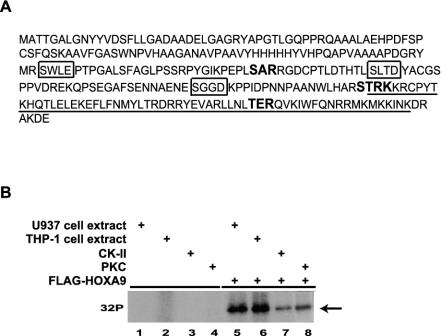
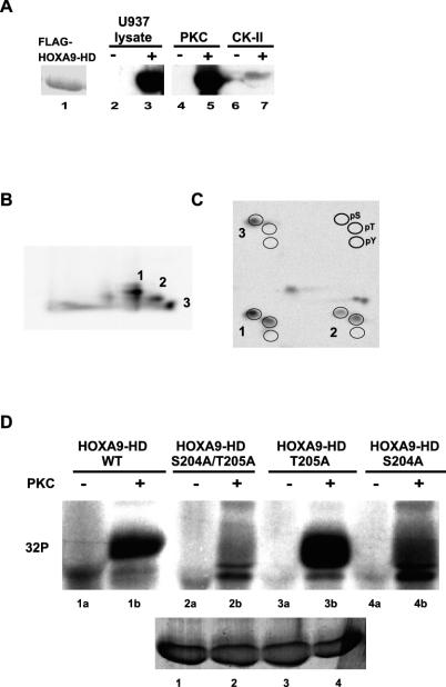
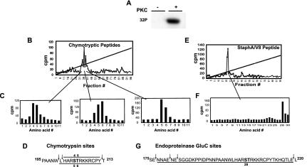
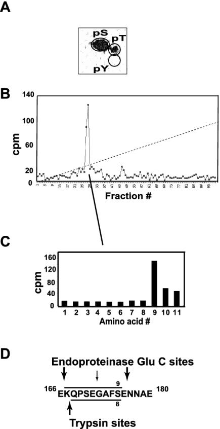
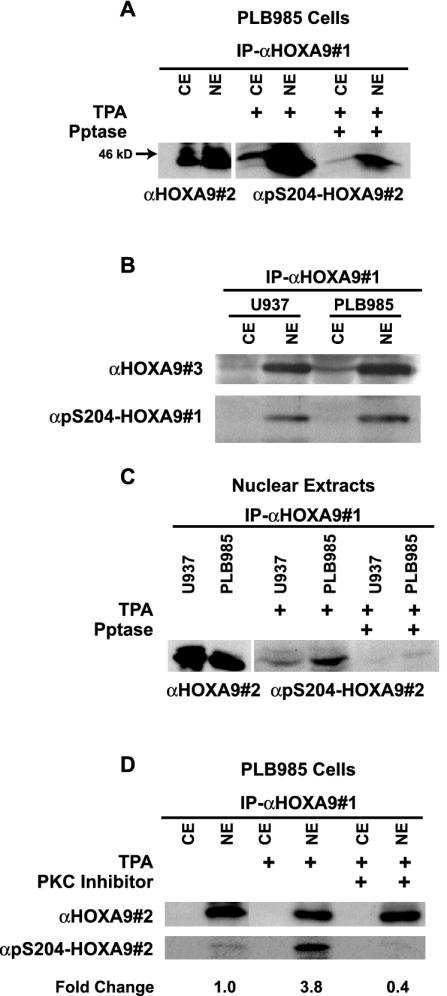
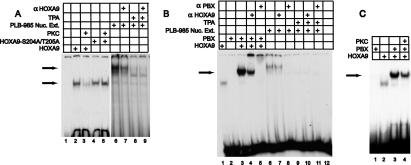
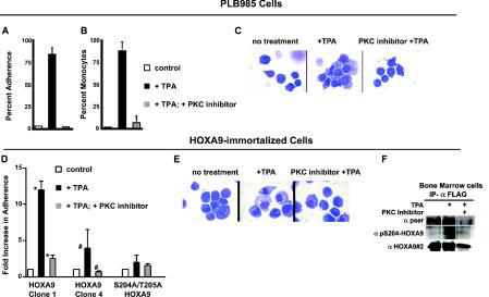
HOXA9 expression is a common feature of acute myeloid leukemia, and high-level expression is correlated with poor prognosis. Moreover, HOXA9 overexpression immortalizes murine marrow progenitors that are arrested at a promyelocytic stage of differentiation when cultured and causes leukemia in recipient mice following transplantation of HOXA9 expressing bone marrow. The molecular mechanisms underlying the physiologic functions and transforming properties of HOXA9 are poorly understood. This study demonstrates that HOXA9 is phosphorylated by protein kinase C (PKC) and casein kinase II and that PKC mediates phosphorylation of purified HOXA9 on S204 as well as on T205, within a highly conserved consensus sequence, in the N-terminal region of the homeodomain. S204 in the endogenous HOXA9 protein was phosphorylated in PLB985 myeloid cells, as well as in HOXA9-immortalized murine marrow cells. This phosphorylation was enhanced by phorbol ester, a known inducer of PKC, and was inhibited by a specific PKC inhibitor. PKC-mediated phosphorylation of S204 decreased HOXA9 DNA binding affinity in vitro and the ability of the endogenous HOXA9 to form cooperative DNA binding complexes with PBX. PKC inhibition significantly reduced the phorbol-ester induced differentiation of the PLB985 hematopoietic cell line as well as HOXA9-immortalized murine bone marrow cells. These data suggest that phorbol ester-induced myeloid differentiation is in part due to PKC-mediated phosphorylation of HOXA9, which decreases the DNA binding of the homeoprotein.


Figures

FIG. 1.
FIG. 1.
HOXA9 is phosphorylated in vitro. (A) Sequence of the HOXA9 full-length protein with the PKC consensus sequences (in boldface type) and CK-II consensus sequences (boxed) as predicted by the Prosite sequence analysis program. The HD sequence is underlined. (B) His-tagged FLAG-HOXA9 protein synthesized using the TNT reticulocyte lysate system or control lysate was subjected to kinase assays with U937 cell extracts, THP-1 cell extracts, purified CK-II, or purified PKC. Radiolabeled bands migrating with the mobility of HOXA9 were detected in the kinase reactions with cell extracts as well as purified enzymes.
FIG. 2.
FIG. 2.
FLAG-HOXA9-HD protein is phosphorylated in vitro by PKC at S204 and T205. (A) FLAG-HOXA9-HD (consisting of the 60-amino-acid HD and 1 N-terminal residue [S204] and 6 C-terminal amino acids [K265-E271]) expressed from the pET28a expression vector was purified as a His-tagged protein on Probond resin and visualized by Coomassie blue staining (lane 1). Lanes 2 to 6 represent autoradiographs of His-tagged FLAG-HOXA9-HD immobilized on the resin (lanes 3, 5, and 7) or control resin that were subjected to kinase assays with U937 cell extracts (lanes 2 and 3), purified PKC (lanes 4 and 5) and purified CK-II (lanes 6 and 7) in the presence of [γ-32P]ATP, followed by elution and electrophoresis. (B) Tryptic peptides of PKC phosphorylated, 32P-labeled FLAG-HOXA9-HD were separated by thin-layer electrophoresis with pH 1.9 buffer in the first dimension and chromatography with n-butanol-pyridine-acetic acid-H2O in the second dimension. Three strong phosphopeptides were detected. (C) Phosphoamino acid analysis was performed on the three phosphopeptides. Phosphorylated serine, threonine, and tyrosine residues migrate in a standard pattern indicated at the top right. (D) Affinity-purified FLAG-HOXA9-HD wild-type (#1) and mutant proteins; S204A/T205A (#2), T205A (#3), S204A (#4), immobilized on beads were subjected to in vitro kinase assay in the absence (lanes 1a to 4a) or presence (lanes 1b to 4b) of purified PKC. Phosphorylated bands for FLAG-HOXA9-HD-WT and -T205A proteins were detected in the PKC-treated reactions, whereas very weak or no phosphorylated bands were detected for FLAG-HOXA9-HD-S204A/T205A and -S204A mutant proteins. The bottom gel represents Coomassie blue-stained bands demonstrating equal amounts of wild-type, and mutant proteins were used for the labeling experiment.
FIG. 3.
FIG. 3.
Identification of PKCα phosphorylation sites in the full-length FLAG-HOXA9 protein. (A) Affinity-purified His-tagged FLAG-HOXA9 was subjected to in vitro kinase assay with [γ-32]ATP in the absence or presence of purified PKC. (B) Chymotryptic peptides of PKCα-phosphorylated 32P-labeled FLAG-HOXA9, were separated by reverse-phase HPLC. (C) The radioactive fractions contributing to each peak were analyzed by Edman degradation. Sequencing of peak 1 released radioactivity in Edman cycles 4 and 5; similarly, sequencing of peaks 2 and 3 released radioactivity in cycles 4, 5, and 6 and cycles 5 and 6, respectively. (D) Sequence of the two overlapping FLAG-HOXA9 chymotryptic peptides, H201-Y212 and L200-Y212, which have serine and threonine residues in positions 4 and 5 and positions 5 and 6, respectively. Peak 1 was generated by chymotryptic peptide H201-Y212, peak 3 was generated by chymotryptic peptide L200-Y212, and peak 2 was generated by a mixture of the two overlapping chymotryptic peptides (E) Similarly, endoproteinase Glu-C peptides of 32P-labeled FLAG-HOXA9 were separated by reverse-phase HPLC and generated a single peak. (F) Edman sequencing of the endoproteinase Glu-C peak released the radioactivity in cycle 28. (G) Sequence of FLAG-HOXA9 with the predicted endoproteinase Glu-C cleavage sites, peptide N177-E219 has S204 at position 28.
FIG. 4.
FIG. 4.
Identification of a single CK-II phosphorylation site in the full-length FLAG-HOXA9 protein. (A) Phosphoamino acid analysis of the CK-II-phosphorylated 32P-labeled His-tagged FLAG-HOXA9 protein indicated that the label was predominantly associated with serine. (B) Endoproteinase Glu-C peptides of CK-II-phosphorylated 32P-labeled FLAG-HOXA9 separated by reverse-phase HPLC generated a single radioactive peak. (C) Edman sequencing of the HPLC radioactive fractions contributing to the peak released the label in cycle 9. (D) Sequence of the FLAG-HOXA9 protein with the predicted endoproteinase Glu-C and trypsin cleavage sites; Endoproteinase Glu-C peptide K167-K176 has S175 at position 9.
FIG. 5.
FIG. 5.
HOXA9 is phosphorylated at S204 in vivo and this phosphorylation is mediated by PKC. (A) Cytoplasmic or nuclear extracts of PLB985 cells were immunoprecipitated with goat α-HOXA9 antibody (αHOXA9#1). Western blot analysis of the immunoprecipitates using affinity-purified chicken α-HOXA9 antibody (αHOXA9#2) showed expression of HOXA9 predominantly in the nuclear extract and to a lesser extent in the cytoplasmic extract. HOXA9 immunoprecipitates from cytoplasmic and nuclear extracts of TPA-stimulated PLB985 cells, treated with or without phosphatase, were probed with α-pS204-HOXA9 antibody (#2). (B) HOXA9 protein from cytoplasmic and nuclear extracts from U937 and PLB985 cells treated with TPA was precipitated with αHOXA9#1 and detected with a rabbit affinity-purified α-HOXA9 antibody (#3) or a different α-pS204-HOXA9 antibody (#1). (C) Immunoprecipitates from nuclear extracts of U937 cells contained HOXA9 protein that was immunoreactive with αHOXA9#2 antibody. HOXA9 immunoprecipitates, from nuclear extracts of TPA-stimulated PLB985 and U937 cells, treated with or without phosphatase, were probed with α-pS204-HOXA9 antibody. (D) PLB985 cells stimulated with either TPA or control were pretreated with or without the PKC inhibitor bisindolylmaleamide1 (5 μM). Cytoplasmic and nuclear extracts were immunoprecipitated with αHOXA9#1, and analyzed by Western blotting with either αHOXA9#2 or α-pS204-HOXA9. Change in HOXA9 phosphorylation level was calculated by NIH ImageQuant analysis.
FIG. 6.
FIG. 6.
Phosphorylated HOXA9 exhibits impaired DNA binding ability. (A) HOXA9-containing EMSA complexes are sensitive to PKC and TPA. EMSA was used to assess DNA binding of in vitro-translated FLAG-HOXA9 and FLAG-HOXA9-S204A/T205A proteins. PKC treatment eliminated HOXA9-DNA binding, but did not affect binding by the mutant protein that could not be phosphorylated on S204 and T205 (compare lanes 3 and 5). The nuclear extracts from PLB985 cells stimulated with TPA that are shown in Fig. 5D were also used for EMSA. These extracts showed reduced EMSA bands compared to controls (compare lanes 6 and 8). The presence of HOXA9 in the EMSA complex formed from PLB985 nuclear extracts was demonstrated using α-HOXA9 antibody (compare lanes 6 and 7). (B) PBX-HOXA9 containing EMSA complexes formed from PLB985 nuclear extracts are sensitive to TPA. The arrow denotes the EMSA complexes formed by HOXA9 and PBX1 (lane 3), which migrate between the band for HOXA9-alone-bound DNA (lane 1) and the band formed by PLB nuclear extract (lane 6). The presence of PBX1 in the extract-derived EMSA band was shown using α-PBX1 antibody (lane 8), while α-HOXA9 antibody partially reduced this complex (lane 7). TPA treatment that induces PKC-mediated phosphorylation of HOXA9 abolishes the EMSA (compare lanes 6 and 9). EMSA was performed using an oligonucleotide probe containing a consensus-binding site for HOXA9 and PBX. (C) In vitro-transcribed and -translated HOXA9 and PBX1a proteins were used for EMSA analysis with a consensus PBX1-HOXA9 target. HOXA9 protein was incubated with PKC buffer control (lane 3) or with PKC (lane 4), prior to EMSA analysis. The arrow marks the migration position of the heterodimeric PBX1a-HOXA9 complex established in previous studies (50), which is reduce by pretreatment of the HOXA9 by PKCα.
FIG. 7.
FIG. 7.
PLB985 and HOXA9-immortalized murine bone marrow cells treated with TPA differentiate into monocytes, and this differentiation is blocked by a PKC inhibitor and appears to require S204/T205 phosphorylation. PLB985 cells (A to C) or HOXA9-immortalized cells (D to F) were treated with dimethyl sulfoxide vehicle or TPA (100 nM) with or without pretreatment with the PKC inhibitor bisindoylmaleamide1 (5 μM) 5 min prior to stimulation with TPA. (A and D) Percent adherence was measured 24 h after addition of TPA. There was a statistically different adherence in the presence of TPA (PLB985 P < 0.001, n = 4) (symbols: *, P < 0.04; #, P < 0.004) but no difference between controls and cells pretreated with inhibitor before TPA treatment. (B) PLB985 differentiation scored on the basis of three monocytic features: nuclear to cytoplasmic ratio, condensed chromatin and cytoplasmic vacuolization. There was a statistically different monocytic differentiation in the presence of TPA (P < 0.001, n = 4) but no difference between controls and cells pretreated with inhibitor before TPA treatment. (C and E) Wright-Giemsa-stained cytospin preparations showing that pretreatment with a PKC inhibitor partially prevented monocytic differentiation of both PLB985 (C) and HOXA9-immortalized cells (E) as reflected by large nuclear size and a smaller cytoplasmic fraction. (F) Endogenous HOXA9 in immortalized marrow line 4 is phosphorylated on S204. Whole-cell lysates were immunoprecipitated with α-FLAG antibody, and the immunoprecipitates were probed with anti-pSer (α-pser), α-pS204-HOXA9, or αHOXA9#2 antibodies. To confirm that S204 and T205 are important for PKC-mediated myeloid cell differentiation, a S204A/T205A HOXA9 protein containing alanines in place of the putative PKC phosphorylation sites was used to immortalize bone marrow cells. The resulting clone showed greatly reduced differentiation response to TPA as measured by cellular adherence (D) or morphology (not shown). Error bars, standard deviations.

Similar articles

Cited by

References

    1. Ausubel, F. M., R. Brent, R. E. Kingston, D. D. Moore, J. G. Seidman, J. A. Smith, and K. Struhl (ed.). 1987. Current protocols in molecular biology, p. 12.1.1-12.1.8. John Wiley & Sons, Hoboken, N.J.
    1. Berry, M., and W. Gehring. 2000. Phosphorylation status of the SCR homeodomain determines its functional activity: essential role for protein phosphatase 2A,B′. EMBO J. 19:2946-2957. - PMC - PubMed
    1. Borrow, J., A. M. Shearman, V. P. J. Stanton, R. Becher, T. Collins, A. J. Williams, I. Dube, F. Katz, Y. L. Kwong, C. Morris, K. Ohyashiki, K. Toyama, J. Rowley, and D. E. Housman. 1996. The t(7;11)(p15;p15) translocation in acute myeloid leukemia fuses the genes for nucleoporin NU98 and class I homeoprotein HOXA9. Nat. Genet. 12:159-167. - PubMed
    1. Bourbon, H. M., E. Martin-Blanco, D. Rosen, and T. B. Kornberg. 1995. Phosphorylation of the Drosophila engrailed protein at a site outside its homeodomain enhances DNA binding. J. Biol. Chem. 270:11130-11139. - PubMed
    1. Boyle, W. J., P. van der Geer, and T. Hunter. 1991. Phosphopeptide mapping and phosphoamino acid analysis by two-dimensional separation on thin-layer cellulose plates. Methods Enzymol. 201:110-143. - PubMed

Publication types

MeSH terms

LinkOut - more resources