. 2004 May;24(9):3838-48.
doi: 10.1128/MCB.24.9.3838-3848.2004.

The NRIF3 family of transcriptional coregulators induces rapid and profound apoptosis in breast cancer cells

Affiliations

The NRIF3 family of transcriptional coregulators induces rapid and profound apoptosis in breast cancer cells

Dangsheng Li et al. Mol Cell Biol. 2004 May.

Abstract

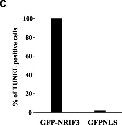
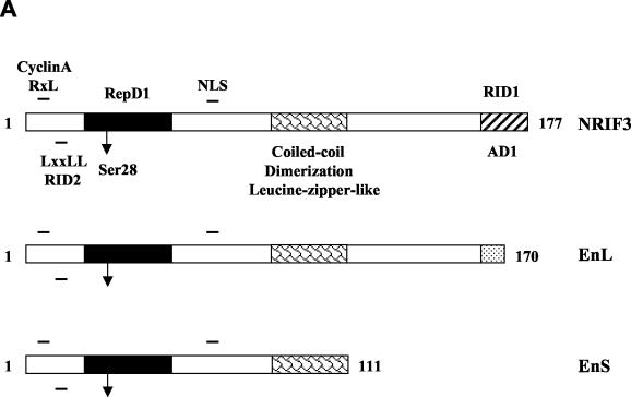
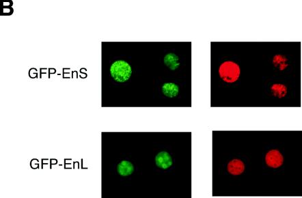
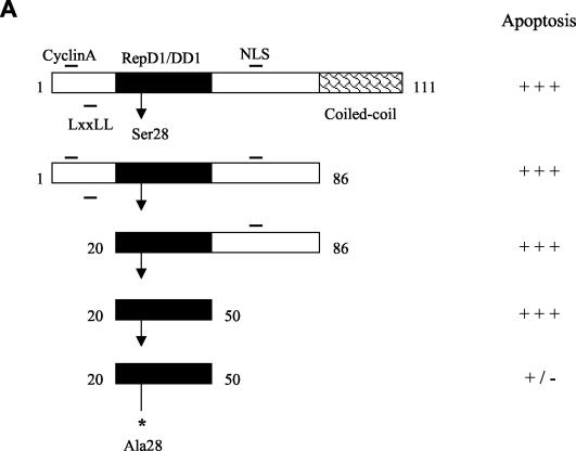
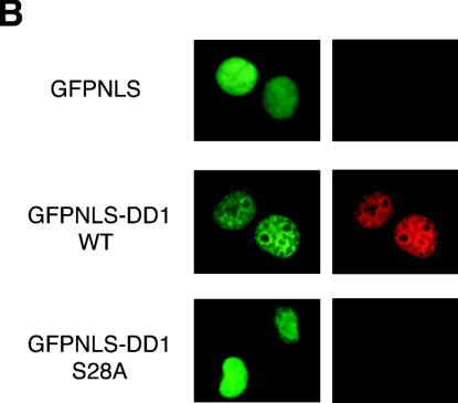
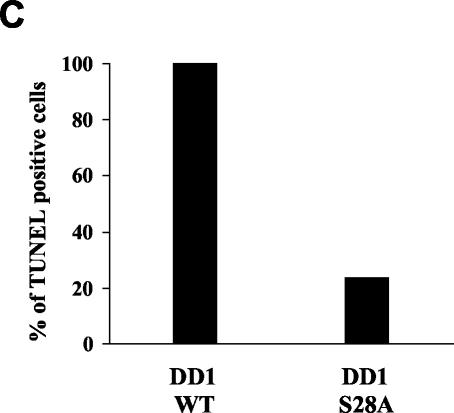
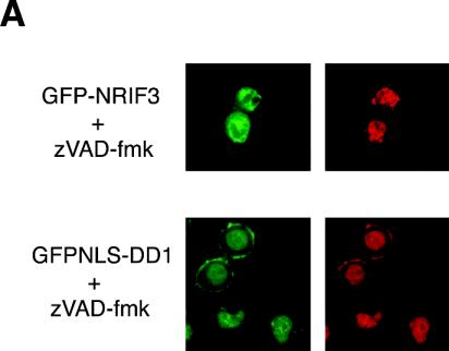
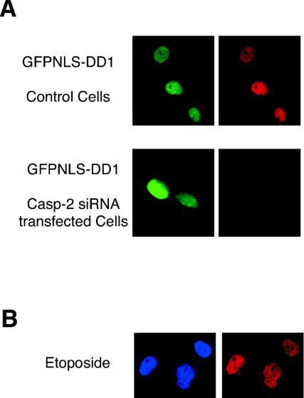
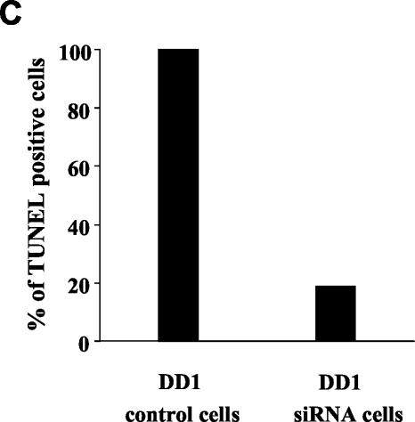
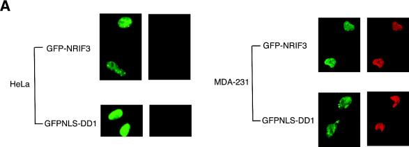
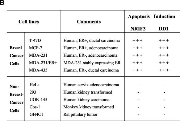
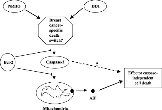
Many anticancer drugs kill cancer cells by inducing apoptosis. Despite the progress in understanding apoptosis, how to harness the cellular death machinery to selectively deliver tumor-specific cytotoxicity (while minimizing damage to other cells) remains an important challenge. We report here that expression of the NRIF3 family of transcriptional coregulators in a variety of breast cancer cell lines induces rapid and profound apoptosis (nearly 100% cell death within 24 h). A novel death domain (DD1) was mapped to a short 30-amino-acid region common to all members of the NRIF3 family. Mechanistic studies showed that DD1-induced apoptosis occurs through a novel caspase 2-mediated pathway that involves mitochondrial membrane permeabilization but does not require other caspases. Interestingly, the cytotoxicity of NRIF3 and DD1 appears to be cell type specific, as they selectively kill breast cancer or related cells but not other examined cells of different origins. Our study demonstrates the feasibility of selectively inducing cytotoxicity in a specific cancer and suggests that breast cancer cells contain a novel "death switch" that can be specifically triggered by NRIF3 or DD1. Strategies utilizing NRIF3 and/or DD1 and/or targeting this death switch may lead to the development of novel and more selective therapeutics against breast cancer.


Figures

FIG. 1.
FIG. 1.
NRIF3 induces apoptosis in T-47D cells. (A) GFP-NRIF3 was expressed in T-47D cells by transient transfection. Twenty-four hours later the green fluorescent cells were collected after sorting by flow cytometry and reinoculated onto coverslips. Cells were then analyzed for apoptosis by annexin V staining (red). Control cells expressing GFP alone were negative for annexin V staining (data not shown). (B) Representative fluorescence micrographs of T-47D cells transfected with either GFPNLS or GFP-NRIF3. Cells were examined for apoptosis by TUNEL assay (red). (C) Quantitative presentation of the experiments in panel B. The percentages of green fluorescent cells that were TUNEL positive were scored for T-47D cells transfected with either GFP-NRIF3 or GFPNLS.
FIG. 1.
FIG. 1.
NRIF3 induces apoptosis in T-47D cells. (A) GFP-NRIF3 was expressed in T-47D cells by transient transfection. Twenty-four hours later the green fluorescent cells were collected after sorting by flow cytometry and reinoculated onto coverslips. Cells were then analyzed for apoptosis by annexin V staining (red). Control cells expressing GFP alone were negative for annexin V staining (data not shown). (B) Representative fluorescence micrographs of T-47D cells transfected with either GFPNLS or GFP-NRIF3. Cells were examined for apoptosis by TUNEL assay (red). (C) Quantitative presentation of the experiments in panel B. The percentages of green fluorescent cells that were TUNEL positive were scored for T-47D cells transfected with either GFP-NRIF3 or GFPNLS.
FIG. 2.
FIG. 2.
Cell death induced by EnS and EnL. (A) Domain organization and functional motifs in NRIF3, EnS, and EnL. NRIF3 contains two nuclear receptor interaction domains (RID1, residues 162 to 177, and RID2, residues 9 to 13) (28, 29). An activation domain (AD1) coresides with RID1. A transrepression domain (RepD1) maps to residues 20 to 50 (29). Also shown are a cyclin A binding motif (RxL; residues 6 to 8), a coiled-coil dimerization domain (residues 86 to 112) that contains a leucine zipper-like motif, and an NLS (residues 63 to 66) (29, 36). Ser28 in RepD1 is marked by an arrow. EnS and EnL share extensive identities with NRIF3 and contain the same domains or motifs except for RID1/AD1. (B) Representative fluorescence micrographs of T-47D cells transfected with either GFP-EnS or GFP-EnL. Cells were examined for apoptosis by TUNEL assay (red).
FIG. 2.
FIG. 2.
Cell death induced by EnS and EnL. (A) Domain organization and functional motifs in NRIF3, EnS, and EnL. NRIF3 contains two nuclear receptor interaction domains (RID1, residues 162 to 177, and RID2, residues 9 to 13) (28, 29). An activation domain (AD1) coresides with RID1. A transrepression domain (RepD1) maps to residues 20 to 50 (29). Also shown are a cyclin A binding motif (RxL; residues 6 to 8), a coiled-coil dimerization domain (residues 86 to 112) that contains a leucine zipper-like motif, and an NLS (residues 63 to 66) (29, 36). Ser28 in RepD1 is marked by an arrow. EnS and EnL share extensive identities with NRIF3 and contain the same domains or motifs except for RID1/AD1. (B) Representative fluorescence micrographs of T-47D cells transfected with either GFP-EnS or GFP-EnL. Cells were examined for apoptosis by TUNEL assay (red).
FIG. 3.
FIG. 3.
NRIF3 contains a novel death domain (DD1). (A) T-47D cells were transfected with each of the indicated constructs expressing various regions of NRIF3 fused to GFP or GFPNLS. Ser28 in wild-type DD1 is marked by an arrow, while Ala28 in the mutant DD1 is marked by an asterisk. These regions were expressed either as a GFP fusion or, for those lacking an intrinsic NLS, as a GFPNLS fusion. Green fluorescent cells were scored for apoptosis by TUNEL assay, annexin V staining, or both. “+++” indicates profound cell death, where nearly 100% of green cells displayed positive staining for TUNEL and/or annexin V, while “−” indicates no apoptosis (less than 2%). (B) Representative fluorescence micrographs of T-47D cells transfected with GFPNLS, wild-type (WT) GFPNLS-DD1 (residues 20 to 50 of NRIF3), or the GFPNLS-DD1 mutant (S28A). Cells were examined for apoptosis by TUNEL assay (red). (C) The Ser28-to-Ala mutation severely compromises the apoptogenic function of DD1. The percentages of green fluorescent cells that were TUNEL positive were scored for T-47D cells transfected with either wild-type (WT) GFPNLS-DD1 or the mutant GFPNLS-DD1S28A.
FIG. 3.
FIG. 3.
NRIF3 contains a novel death domain (DD1). (A) T-47D cells were transfected with each of the indicated constructs expressing various regions of NRIF3 fused to GFP or GFPNLS. Ser28 in wild-type DD1 is marked by an arrow, while Ala28 in the mutant DD1 is marked by an asterisk. These regions were expressed either as a GFP fusion or, for those lacking an intrinsic NLS, as a GFPNLS fusion. Green fluorescent cells were scored for apoptosis by TUNEL assay, annexin V staining, or both. “+++” indicates profound cell death, where nearly 100% of green cells displayed positive staining for TUNEL and/or annexin V, while “−” indicates no apoptosis (less than 2%). (B) Representative fluorescence micrographs of T-47D cells transfected with GFPNLS, wild-type (WT) GFPNLS-DD1 (residues 20 to 50 of NRIF3), or the GFPNLS-DD1 mutant (S28A). Cells were examined for apoptosis by TUNEL assay (red). (C) The Ser28-to-Ala mutation severely compromises the apoptogenic function of DD1. The percentages of green fluorescent cells that were TUNEL positive were scored for T-47D cells transfected with either wild-type (WT) GFPNLS-DD1 or the mutant GFPNLS-DD1S28A.
FIG. 4.
FIG. 4.
Cell death mediated by NRIF3 or DD1 is insensitive to zVAD-fmk. (A) T-47D cells were transfected with either GFP-NRIF3 or GFPNLS-DD1 in the absence or presence of the broad-spectrum caspase inhibitor zVAD-fmk. When present, the inhibitor was incubated with the cells before and after transfection. Cells were examined for apoptosis by TUNEL assay (red). Representative fluorescence micrographs are shown for cells treated with zVAD-fmk. (B) Quantitative presentation of the experiments in panel A. The percentages of green fluorescent cells that were TUNEL positive were scored for T-47D cells transfected with either GFP-NRIF3 or GFPNLS-DD1 in the absence or presence of zVAD-fmk.
FIG. 4.
FIG. 4.
Cell death mediated by NRIF3 or DD1 is insensitive to zVAD-fmk. (A) T-47D cells were transfected with either GFP-NRIF3 or GFPNLS-DD1 in the absence or presence of the broad-spectrum caspase inhibitor zVAD-fmk. When present, the inhibitor was incubated with the cells before and after transfection. Cells were examined for apoptosis by TUNEL assay (red). Representative fluorescence micrographs are shown for cells treated with zVAD-fmk. (B) Quantitative presentation of the experiments in panel A. The percentages of green fluorescent cells that were TUNEL positive were scored for T-47D cells transfected with either GFP-NRIF3 or GFPNLS-DD1 in the absence or presence of zVAD-fmk.
FIG. 5.
FIG. 5.
DD1-mediated cell death involves MMP. (A) Representative fluorescence micrographs of T-47D cells transfected either with GFPNLS-DD1 or with GFPNLS-DD1 and Bcl-2. Cells were examined for apoptosis by TUNEL assay (red). (B) Quantitative presentation of the experiments in panel A. The percentages of green fluorescent cells that were TUNEL positive were scored for T-47D cells transfected either with GFPNLS-DD1 or with GFPNLS-DD1 and Bcl-2. (C) T-47D cells were transfected to express AIF-GFP along with either a control vector or a vector expressing DD1. Approximately 5 h after transfection the cells were fixed and subjected to TUNEL assay. The cells were then examined by fluorescence microscopy for subcellular localization of AIF-GFP (green) and for apoptosis (red). Nuclei were stained with Hoechst dye (blue). Representative fluorescence micrographs of cells transfected with the control vector or DD1 are compared.
FIG. 5.
FIG. 5.
DD1-mediated cell death involves MMP. (A) Representative fluorescence micrographs of T-47D cells transfected either with GFPNLS-DD1 or with GFPNLS-DD1 and Bcl-2. Cells were examined for apoptosis by TUNEL assay (red). (B) Quantitative presentation of the experiments in panel A. The percentages of green fluorescent cells that were TUNEL positive were scored for T-47D cells transfected either with GFPNLS-DD1 or with GFPNLS-DD1 and Bcl-2. (C) T-47D cells were transfected to express AIF-GFP along with either a control vector or a vector expressing DD1. Approximately 5 h after transfection the cells were fixed and subjected to TUNEL assay. The cells were then examined by fluorescence microscopy for subcellular localization of AIF-GFP (green) and for apoptosis (red). Nuclei were stained with Hoechst dye (blue). Representative fluorescence micrographs of cells transfected with the control vector or DD1 are compared.
FIG. 6.
FIG. 6.
Requirement for caspase 2 in DD1-mediated apoptosis. (A) T-47D cells pretreated with caspase 2 siRNA or mock-treated control cells were transfected with GFPNLS-DD1. Cells were examined for apoptosis by TUNEL assay (red). Representative fluorescence micrographs of siRNA-treated or mock-treated cells are compared. (B) T-47D cells pretreated with caspase 2 siRNA or mock-treated control cells were incubated with etoposide. Cells were examined for apoptosis by TUNEL assay (red), while the nuclei were visualized by Hoechst staining (blue). Representative fluorescence micrographs are shown for the siRNA-treated cells. Similar results were observed for the mock-treated control cells (data not shown). (C) Quantitative presentation of the experiments in panel A. The percentages of green fluorescent cells (expressing GFPNLS-DD1) that were TUNEL positive were scored for cells pretreated with caspase 2 siRNA or for mock-treated control cells.
FIG. 6.
FIG. 6.
Requirement for caspase 2 in DD1-mediated apoptosis. (A) T-47D cells pretreated with caspase 2 siRNA or mock-treated control cells were transfected with GFPNLS-DD1. Cells were examined for apoptosis by TUNEL assay (red). Representative fluorescence micrographs of siRNA-treated or mock-treated cells are compared. (B) T-47D cells pretreated with caspase 2 siRNA or mock-treated control cells were incubated with etoposide. Cells were examined for apoptosis by TUNEL assay (red), while the nuclei were visualized by Hoechst staining (blue). Representative fluorescence micrographs are shown for the siRNA-treated cells. Similar results were observed for the mock-treated control cells (data not shown). (C) Quantitative presentation of the experiments in panel A. The percentages of green fluorescent cells (expressing GFPNLS-DD1) that were TUNEL positive were scored for cells pretreated with caspase 2 siRNA or for mock-treated control cells.
FIG. 7.
FIG. 7.
Cell type specificity in cytotoxicity mediated by NRIF3 and DD1. (A) Various breast cancer cell lines and other cells were transfected with either GFP-NRIF3 or GFPNLS-DD1. Cells were examined for apoptosis by TUNEL assay (red). Representative fluorescence micrographs of transfected HeLa and MDA-MB-231 cells are compared. (B) Summary of results from all cell lines examined in panel A. “+++” indicates profound apoptosis (>90% of green fluorescent cells were TUNEL positive) while “−” indicates no apoptosis (less than 2% positive).
FIG. 7.
FIG. 7.
Cell type specificity in cytotoxicity mediated by NRIF3 and DD1. (A) Various breast cancer cell lines and other cells were transfected with either GFP-NRIF3 or GFPNLS-DD1. Cells were examined for apoptosis by TUNEL assay (red). Representative fluorescence micrographs of transfected HeLa and MDA-MB-231 cells are compared. (B) Summary of results from all cell lines examined in panel A. “+++” indicates profound apoptosis (>90% of green fluorescent cells were TUNEL positive) while “−” indicates no apoptosis (less than 2% positive).
FIG. 8.
FIG. 8.
Model for NRIF3- or DD1-induced apoptosis. We suggest that breast cancer cells contain a specific death switch that can be selectively triggered by NRIF3 or its death domain DD1. Triggering of this switch by NRIF3 or DD1 leads to activation of caspase 2. Activated caspase 2 promotes MMP, which results in the release of AIF. The released AIF then mediates effector caspase-independent cell death (which is insensitive to zVAD-fmk). The antiapoptotic factor Bcl-2 could inhibit this pathway by acting either upstream of caspase 2 (to prevent its activation) or downstream of caspase 2 (to prevent caspase 2-mediated MMP) (24). It is also possible that activated caspase 2 can directly elicit cell death (dashed line) in addition to the depicted mitochondrion-mediated pathway.

References

    1. Adam, S. A., and L. Gerace. 1991. Cytosolic proteins that specifically bind nuclear location signals are receptors for nuclear import. Cell 66:837-847. - PubMed
    1. Afonja, A., B. M. Raaka, A. Huang, S. Das, X. Zhao, E. Helmer, D. Juste, and H. H. Samuels. 2002. RAR agonists stimulate SOX9 gene expression in breast cancer cell lines: evidence for a role in retinoid-mediated growth inhibition. Oncogene 21:7850-7860. - PubMed
    1. Antonsson, B. 2001. Bax and other pro-apoptotic Bcl-2 family “killer-proteins” and their victim the mitochondrion. Cell Tissue Res. 306:347-361. - PubMed
    1. Baliga, B. C., P. A. Colussi, S. H. Read, M. M. Dias, D. A. Jans, and S. Kumar. 2003. Role of prodomain in importin-mediated nuclear localization and activation of caspase-2. J. Biol. Chem. 278:4899-4905. - PubMed
    1. Benson, J. R., and V. Pitsinis. 2003. Update on clinical role of tamoxifen. Curr. Opin. Obstet. Gynecol. 15:13-23. - PubMed

Publication types

MeSH terms