. 2004 May;24(9):3957-71.
doi: 10.1128/MCB.24.9.3957-3971.2004.

Human enhancer of invasion-cluster, a coiled-coil protein required for passage through mitosis

Affiliations

Human enhancer of invasion-cluster, a coiled-coil protein required for passage through mitosis

Margret B Einarson et al. Mol Cell Biol. 2004 May.

Abstract

In a cross-species overexpression approach, we used the pseudohyphal transition of Saccharomyces cerevisiae as a model screening system to identify human genes that regulate cell morphology and the cell cycle. Human enhancer of invasion-cluster (HEI-C), encoding a novel evolutionarily conserved coiled-coil protein, was isolated in a screen for human genes that induce agar invasion in S. cerevisiae. In human cells, HEI-C is primarily localized to the spindle during mitosis. Depletion of HEI-C in vivo with short interfering RNAs results in severe mitotic defects. Analysis by immunofluorescence, flow cytometry analysis, and videomicroscopy indicates that HEI-C-depleted cells form metaphase plates with normal timing after G(2)/M transition, although in many cases cells have disorganized mitotic spindles. Subsequently, severe defects occur at the metaphase-anaphase transition, characterized by a significant delay at this stage or, more commonly, cellular disintegration accompanied by the display of classic biochemical markers of apoptosis. These mitotic defects occur in spite of the fact that HEI-C-depleted cells retain functional cell cycle checkpoints, as these cells arrest normally following nocodazole or hydroxyurea treatment. These results place HEI-C as a novel regulator of spindle function and integrity during the metaphase-anaphase transition.


Figures
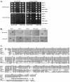
FIG. 1.
FIG. 1.
HEI-C causes invasive and filamentous growth in S. cerevisiae. (A) Assay for invasive growth. Diploid CGx75 or isogenic haploid CG188 S. cerevisiae strains were transformed with plasmids isolated in the library screen expressing HEF1 (amino acids 660 to 834), PRK2 (amino acids 505 to 984), a kinase dead mutant of the PRK2 clone (PRK2-KD) or HEI-C (amino acids 1 to 278, full-length), and two independent transformants replica plated onto plates lacking uracil and tryptophan and with glucose (gene expression off) or lacking uracil and tryptophan and with galactose (gene expression on). After 24 h of growth, the plates were photographed (prewash), washed with running distilled water, and rephotographed (postwash). (B) Assay of filamentous growth. Diploid CGx75 or isogenic haploid CG188 S. cerevisiae strains were transformed with the HEF1, PRK2, PRK2.KD, or HEI-C clones isolated in the invasive screen and streaked onto SLAGR low-nitrogen medium poured on glass slides. After 24 h of growth, the filamentous phenotype was recorded. (C) Comparison of human HEI-C to rat HEI-C and of protein sequences translated from a compiled mouse EST clone, a bovine EST, a compiled chicken EST, a frog EST, and a compiled zebra fish EST demonstrates sequence homology. Identical residues are shaded and boxed; the bottom line of the sequence is the consensus.
FIG. 2.
FIG. 2.
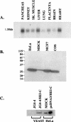
Expression of HEI-C mRNA and protein. (A) A multiple-tissue Northern blot was probed with a 32P-labeled HEI-C oligonucleotide probe. A single message of approximately 1.3 kb was detected. (B) Western analysis of 10 μg of total-cell lysates from HeLa, MDCK, MCF7, and COS7 cells. Cells were lysed in RIPA buffer, subjected to electrophoresis on an SDS-10% PAGE gel, transferred to a polyvinylidene difluoride membrane, and immunoblotted with affinity-purified rabbit polyclonal antipeptide HEI-C antibodies. (C) Expression of the HEI-C clone in S. cerevisiae and mammalian cells produces a protein that comigrates with endogenous HEI-C. Total-cell lysate isolated from CGx75 transformed with vector alone (lane 1) or HEI-15 (lane 2) and grown in galactose and total-cell lysate from mock-transfected HeLa cells (lane 3) or HeLa cells transfected with HEI-C (lane 4) were subjected to electrophoresis on an SDS-10% PAGE gel, transferred to a polyvinylidene difluoride membrane, and probed with affinity-purified anti-peptide HEI-C antibodies.
FIG. 3.
FIG. 3.
HEI-C localizes to the spindle during mitosis. (A) MCF7 cells were initially synchronized with nocodazole, released, and allowed to progress through mitosis. HEI-C is shown in green. DNA was labeled with propidium iodide and digitally assigned to be blue. Bar, 5 μm. (B) Confocal analysis of unsynchronized cells double-labeled for HEI-C (in green) and α-tubulin (in red). (C) Interphase MCF7 cells labeled to visualize HEI-C (green), γ-tubulin (red), or DNA (blue). The inset shows a zoomed yellow colocalization of HEI-C and γ-tubulin only (e.g., in the absence of the blue channel). (D) MDCK cells were stained with antibody to HEI-C (green) or to E-cadherin (red) to demonstrate HEI-C localization to cell junctions. (E) Western blot analysis of HeLa cells synchronized by double thymidine block (time zero), released, then harvested at 4, 6, 8, 10, or 12 h, as indicated, and compared with asynchronous cells (AS). Lysates were subjected to electrophoresis on an SDS-10% PAGE gel, transferred to a polyvinylidene difluoride membrane, and probed with affinity-purified anti-peptide HEI-C antibodies.
FIG. 4.
FIG. 4.
HEI-C is associated in vitro with mitotic asters. (A) HeLa cells were lysed by Dounce homogenization in PHEM buffer, and the cell lysate was precleared by centrifugation. The supernatant was incubated either with (+MT) or without (-MT) microtubules that had been previously polymerized from purified tubulin by incubation in G-PEM buffer. The reactions were layered on a 15% sucrose solution and centrifuged. Total-cell lysate (T) or the resulting supernatant (S) or pellet (P) fractions were subjected to SDS-PAGE, and proteins were subsequently transferred to polyvinylidene difluoride and immunoblotted with affinity-purified anti-HEI-C antibodies. Quantitation was done with NIH Image analysis of scanned films. (B) Taxol and ATP were added to HeLa cell mitotic extracts to stimulate aster formation. Asters were pelleted, and the insoluble (P) and soluble (S) fractions were subjected to electrophoresis and immunoblotting with the indicated antibodies and quantitation as in A. (C) A HeLa cell mitotic extract was fractionated on a 5 to 20% sucrose gradient, and fractions ranging from the top (no. 1) to bottom (no. 14) were collected. Western analysis of these fractions was performed with antibodies specific for HEI-C, dynactin, and tubulin, as indicated.
FIG.5.
FIG.5.
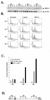
Decrease in G2/M compartment of the cell cycle is accompanied by the appearance of a sub-G1 population in HEI-C-depleted cells. (A) Western analysis of siRNA-transfected cells. HeLa cells were untreated (U), treated with transfection reagent alone (M), transfected with a control siRNA (C), or transfected with an siRNA directed against HEI-C, HEIC.A (A). Cell lysates were subjected to electrophoresis, transferred to a polyvinylidene difluoride membrane, probed with antibody to HEI-C antibodies, and subsequently stripped and reprobed with actin as a loading control. (B) HeLa cells that were untreated, transfected with a control siRNA, or transfected with the HEIC.A siRNA were harvested at the indicated number of days (d) after siRNA transfection and fixed at −20°C in 70% ethanol. The cells were subsequently stained with propidium iodide and analyzed by FACScan. (C) HeLa cells were either left untreated or transfected with either a control siRNA or the HEI-C-directed siRNA HEIC.A and analyzed simultaneously for live annexin and propidium iodide staining at the times indicated after transfection by FACScan. Cell populations were analyzed with FlowJo software. The percent annexin-positive cells is shown. (D) Western analysis of HeLa cells untreated (U), transfected with a control siRNA (C), or transfected with the HEI-C-directed siRNA HEIC.A (A). Cells were lysed in RIPA buffer, subjected to electrophoresis on an SDS-10% PAGE gel, transferred to a polyvinylidene difluoride membrane, and immunoblotted with anti-p85 PARP antibodies.
FIG. 6.
FIG. 6.
HEI-C is required for passage through mitosis. (A) HeLa cells were either left untreated, transfected with a control siRNA, or transfected with the HEIC.A siRNA for 72 h. The cells were either allowed to continue untreated (asynchronous, AS) or treated with hydroxyurea for 20 h prior to harvest (i.e., at 52 h following siRNA transfection) to block cell cycle progression (+HU, 0) or treated with hydroxyurea, and then washed free of drug and released to enter the cell cycle for the indicated number of hours (6, 8, or 10 h postrelease [REL]). Cells were harvested and fixed at −20°C in 70% ethanol. The cells were subsequently stained with propidium iodide and analyzed by FACScan. (B) Western analysis of HeLa cells untreated (U), transfected with a control siRNA (C), or transfected with the HEIC-directed siRNA, HEIC.A (A). Cells were lysed in RIPA buffer, subjected to electrophoresis on an SDS-10% PAGE gel, transferred to a polyvinylidene difluoride membrane, and immunoblotted with affinity-purified rabbit polyclonal anti-PARP antibodies.
FIG. 7.
FIG. 7.
Video microscopy of HEI-C-depleted cells in mitosis. HeLa cells were either transfected with the HEIC.A siRNA (panels A to H) or transfected with a control siRNA (panels I to L) for 72 h. 20 h prior to assessment, cells were treated with hydroxyurea to block cell cycle progression, then washed free of drug, and allowed to enter the cell cycle to enrich for cells in M phase. The first frame corresponds to the first appearance of a metaphase plate, while the times shown on each frame indicate time since the first frame. Asterisks mark cells tracked for the indicated time intervals.
FIG. 8.
FIG. 8.
Confocal analysis of HEI-C-depleted cells. HeLa cells that were depleted of HEIC.A (a to f) versus nondepleted controls (g and h) were fixed in 4% paraformaldehyde and costained with antitubulin antibodies (A) and SYTOX-Green nucleic acid stain (B). (C) Merged images of tubulin in red and DNA in blue. Bar, 10 μm.
FIG. 9.
FIG. 9.
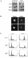
HEI-C is not required for spindle assembly, and HEI-C depletion does not impair the spindle checkpoint. (A) HeLa cell mitotic extract was immunodepleted with protein A-conjugated agarose containing immunoglobulin G from the preimmune rabbit serum (A) or protein A-conjugated agarose containing affinity-purified antibodies against HEI-C (B). The protein A-conjugated agarose was recovered (PAb) from the depletion steps, and the remainder of the extracts was separated into soluble (S) and insoluble (P) fractions following aster assembly by centrifugation at 10,000 × g. These fractions were subjected to Western analysis (D) with antibodies to HEI-C, NuMA, and tubulin as indicated. (B) Following depletion as described for A., microtubule asters were assembled under standard conditions with taxol and ATP for 30 to 60 min at 30°C and processed for indirect immunofluorescence (A and B) with α-tubulin- and NuMA-specific antibodies as indicated. (C) HEI-C-depleted cells have an intact spindle checkpoint. HeLa cells were either untreated, transfected with a control siRNA, or transfected with the HEIC.A siRNA for 48 h, then treated with nocodazole (+nocodazole) for 14 h or left untreated (−nocodazole). Cells were harvested by fixation at −20°C in 70% ethanol. The cells were subsequently stained with propidium iodide and analyzed by FACScan.

References

    1. Amon, A. 1999. The spindle checkpoint. Curr. Opin. Genet. Dev. 9:69-75. - PubMed
    1. Barral, Y., S. Jentsch, and C. Mann. 1995. G1 cyclin turnover and nutrient uptake are controlled by a common pathway in yeast. Genes Dev. 9:399-409. - PubMed
    1. Braverman, L. E., and L. A. Quilliam. 1999. Identification of Grb4/Nckbeta, a src homology 2 and 3 domain-containing adapter protein having similar binding and biological properties to Nck. J. Biol. Chem. 274:5542-5549. - PubMed
    1. Burns, T. F., P. Fei, K. A. Scata, D. T. Dicker, and W. S. El-Deiry. 2003. Silencing of the novel p53 target gene Snk/Plk2 leads to mitotic catastrophe in paclitaxel (taxol)-exposed cells. Mol. Cell. Biol. 23:5556-5571. - PMC - PubMed
    1. Chan, G. K., and T. J. Yen. 2003. The mitotic checkpoint: a signaling pathway that allows a single unattached kinetochore to inhibit mitotic exit. Prog. Cell Cycle Res. 5:431-439. - PubMed

Publication types

MeSH terms

LinkOut - more resources