. 2004 May;24(9):3992-4003.
doi: 10.1128/MCB.24.9.3992-4003.2004.

Acquired expression of periostin by human breast cancers promotes tumor angiogenesis through up-regulation of vascular endothelial growth factor receptor 2 expression

Affiliations

Acquired expression of periostin by human breast cancers promotes tumor angiogenesis through up-regulation of vascular endothelial growth factor receptor 2 expression

Rong Shao et al. Mol Cell Biol. 2004 May.

Abstract

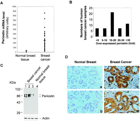
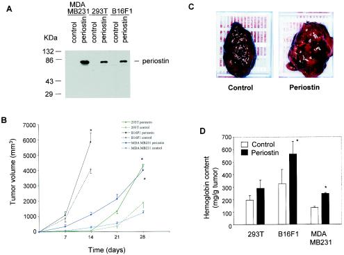
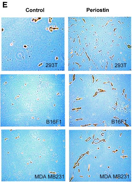
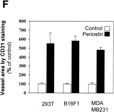
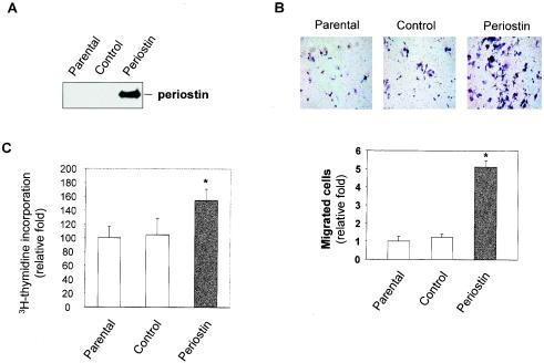
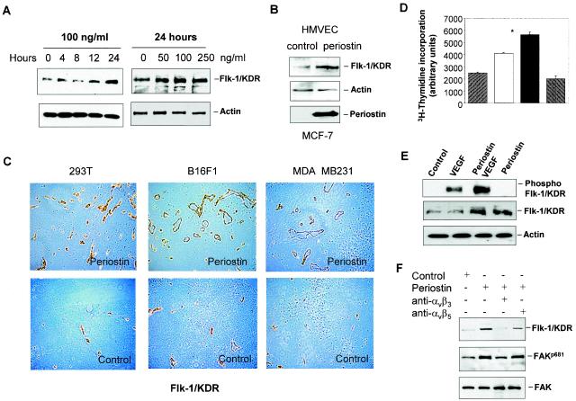
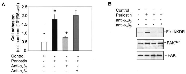
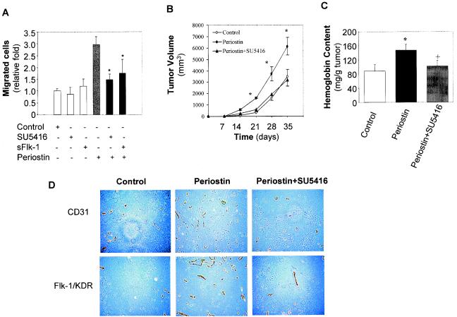
The late stages of human breast cancer development are poorly understood complex processes associated with the expression of genes by cancers that promote specific tumorigenic activities, such as angiogenesis. Here, we describe the identification of periostin as a mesenchyme-specific gene whose acquired expression by human breast cancers leads to a significant enhancement in tumor progression and angiogenesis. Undetectable in normal human breast tissues, periostin was found to be overexpressed by the vast majority of human primary breast cancers examined. Tumor cell lines engineered to overexpress periostin showed a phenotype of accelerated growth and angiogenesis as xenografts in immunocompromised animals. The underlying mechanism of periostin-mediated induction of angiogenesis was found to derive in part from the up-regulation of the vascular endothelial growth factor receptor Flk-1/KDR by endothelial cells through an integrin alpha(v)beta(3)-focal adhesion kinase-mediated signaling pathway. These findings demonstrate the presence of a novel mechanism by which tumor angiogenesis is acquired with the expression of a mesenchyme-specific gene as a crucial step in late stages of tumorigenesis.


Figures

FIG. 1.
FIG. 1.
High levels of periostin expression are associated with human breast cancers. (A) Periostin expression pattern based on gene array data. The raw data from gene array analysis of the expression of periostin in normal (3 samples) and breast cancer (50 samples) tissues are plotted (the mean value for normal tissues at 106 as a baseline versus that for breast cancer samples at 2,100). (B) The same gene array data on periostin expression by 50 breast cancers were categorized into different groups based on levels of periostin overexpression. (C) Periostin protein expression in normal and tumor tissue samples. Tissue extracts from normal or breast cancer tissue samples were subjected to immunoblot analysis with a polyclonal antiperiostin antibody. The results shown in lanes 1 and 2 are from tumor samples from the group with the highest levels of periostin mRNA expression (>30-fold). The result shown in lane 3 was from tumor tissue with less than a fivefold increase in periostin mRNA expression. The higher band of the doublet is likely the unprocessed form of periostin, and the lower band is likely the secreted form of periostin with its signal sequence cleaved from the N terminus. (D) Tissue sections from human normal breast and breast cancer samples were analyzed by immunohistochemical staining and representative sections of each type of sample are shown. Red immunostaining represents positive staining for periostin protein in tumor tissues but not in normal tissues (magnification, ×200).
FIG. 2.
FIG. 2.
Periostin enhances tumor growth and angiogenesis of xenografts in immunocompromised mice. (A) Generation of periostin-producing cells. The production of periostin by cell populations from 293T, B16F1, and MDA-MB-231 cell lines was examined by immunoblotting with conditioned culture media. (B) Time course of tumor growth. The growth rate of tumors derived from periostin-producing or control 293T, B16F1, or MDA-MB-231 cells was monitored weekly by the measurement of tumor volume. (C) Higher levels of hemorrhage were associated with tumors derived from periostin-producing cells than from control cells. As an example, a pair of tumors derived from B16F1 cells was photographed. (D) Higher levels of hemoglobin content were associated with tumors derived from periostin-producing cells. *, P was <0.05 in a comparison with control groups. (E) Higher levels of CD31 expression were detected in tumors derived from periostin-producing cells. Tumor sections were stained with an anti-CD31 antibody, and representative sections for each type of sample are shown. Brown staining distributed along the vasculature network represents CD31-positive endothelial cells (magnification, ×200). (F) Vessel area was determined by quantitation of CD31-positive staining in fixed-size areas of tumor sections by using an image-scanning program developed by the National Institutes of Health.
FIG. 2.
FIG. 2.
Periostin enhances tumor growth and angiogenesis of xenografts in immunocompromised mice. (A) Generation of periostin-producing cells. The production of periostin by cell populations from 293T, B16F1, and MDA-MB-231 cell lines was examined by immunoblotting with conditioned culture media. (B) Time course of tumor growth. The growth rate of tumors derived from periostin-producing or control 293T, B16F1, or MDA-MB-231 cells was monitored weekly by the measurement of tumor volume. (C) Higher levels of hemorrhage were associated with tumors derived from periostin-producing cells than from control cells. As an example, a pair of tumors derived from B16F1 cells was photographed. (D) Higher levels of hemoglobin content were associated with tumors derived from periostin-producing cells. *, P was <0.05 in a comparison with control groups. (E) Higher levels of CD31 expression were detected in tumors derived from periostin-producing cells. Tumor sections were stained with an anti-CD31 antibody, and representative sections for each type of sample are shown. Brown staining distributed along the vasculature network represents CD31-positive endothelial cells (magnification, ×200). (F) Vessel area was determined by quantitation of CD31-positive staining in fixed-size areas of tumor sections by using an image-scanning program developed by the National Institutes of Health.
FIG. 3.
FIG. 3.
Periostin secreted by tumor cells promotes angiogenic activities of HMVEC. (A) The conditioned media from parental, vector-transfected, or periostin-producing MCF-7 cells were collected, and periostin expression was determined by immunoblotting. (B) Tumor cell-derived periostin enhances HMVEC migration. The three types of cells indicated in panel A were grown in serum-free media for 48 h in the lower chamber of transwells. HMVEC were transferred onto the upper chamber, and trapped cells were counted 4 h later. (C) Tumor cell-derived periostin enhances proliferation of HMVEC. HMVEC were incubated with conditioned media derived from the three types of cells described in panel A for 12 h, and [3H]thymidine incorporation was measured following a labeling period of 6 h. *, P was <0.05 compared with parental or control cells.
FIG. 4.
FIG. 4.
Up-regulation of Flk-1/KDR is at least in part responsible for periostin-induced angiogenesis. (A) Recombinant periostin induces the expression of Flk-1/KDR in a time- and dose-dependent manner. HMVEC were treated with periostin (100 ng/ml) for up to 24 h or treated with different concentrations of periostin for 24 h. Protein samples from the treated cells were analyzed for Flk-1/KDR expression by immunoblotting. Actin was used as a loading control. (B) Periostin derived from conditioned medium of MCF-7 cells induces Flk-1/KDR expression. Control or periostin-producing MCF-7 cells shown at the bottom panel were incubated with serum-free medium for 48 h. HMVEC were incubated with the conditioned media for 24 h prior to analysis for Flk-1/KDR expression. (C) Increased presence of Flk-1/KDR associated with blood vessels is detected in tumors derived from periostin-producing cells. Tumor sections derived from 293T, B16F1, or MDA-MB-231 cells were subjected to immunohistochemical staining analysis with an anti-Flk-1/KDR antibody. (D) Periostin pretreatment potentiates HMVEC proliferation in response to VEGF. HMVEC were pretreated with periostin (100 ng/ml) for 12 h. After the cells were washed, VEGF (10 ng/ml) was added for 12 h to determine the effect on cell proliferation. *, P was <0.05 compared with VEGF treatment alone. (E) Periostin pretreatment potentiates Flk-1/KDR phosphorylation in response to VEGF. The same assay condition was used as described above except that the incubation time for VEGF was 5 min, followed by the quantitative determination of phosphorylated Flk-1/KDR by immunoblotting with a specific anti-phospho-Tyr 951 antibody.
FIG. 5.
FIG. 5.
The αvβ3 integrin-FAK signaling pathway mediates the induction of Flk-1/KDR expression. (A) HMVEC (5 × 10⁴ cells/well) were cultured in a 96-well plate precoated with periostin (10 μg/ml) in the presence of anti-αvβ3 or anti-αvβ5 integrin antibody (10 μg/ml) for 1 h (noncoated wells were used as the control). Adhesive cells were counted following washing. *, P was <0.05 compared with the control cells; +, P was <0.05 compared with the cells plated on periostin-coated wells alone. (B) in the presence or absence of the indicated specific anti-integrin antibodies, HMVEC were treated with periostin (100 ng/ml) for 12 h to examine the effect on the up-regulation of Flk-1/KDR as determined by Western blotting. Alternatively, the same assay conditions were used except that the time of incubation was reduced to 15 min to observe a change in FAK autophosphorylation on Tyr 681 (FAKp681). An antibody against FAK was used as a loading control.
FIG. 6.
FIG. 6.
Inhibition of Flk-1/KDR abolished periostin-induced enhancement in tumor growth and angiogenesis. (A) Inhibition of Flk-1/KDR blocks periostin-induced cell migration. HMVEC were employed for a migration assay in the presence of SU5416 (20 μM), sFlk-1 (100 ng/ml), periostin (100 ng/ml), or different combinations as indicated. *, P was <0.05 compared with the group treated with periostin alone. (B) The enhanced tumor growth by periostin-producing 293T cells was completely reversed by the Flk-1/KDR inhibitor SU5416. The time course of 293T tumor growth as measured by tumor volume was plotted with control, periostin-producing cells, or periostin plus SU5416 as described in Materials and Methods. *, P was <0.05 compared with control or periostin plus SU5416. (C and D) Reduced tumor growth in the presence of SU5416 was correlated with a reduction in angiogenesis. Hemoglobin content in tumors was measured as previously described. *, P was <0.05 compared with control; +, P was <0.05 compared with periostin. Tumor sections were immunohistochemically stained with anti-CD31 and Flk-1/KDR antibodies, and representative sections of each type of sample are shown (magnification, ×200).

References

    1. Abu-Ghazaleh, R., J. Kabir, H. Jia, M. Lobo, and I. Zachary. 2001. Src mediates stimulation by vascular endothelial growth factor of the phosphorylation of focal adhesion kinase at tyrosine 861, and migration and anti-apoptosis in endothelial cells. Biochem. J. 360:255-264. - PMC - PubMed
    1. Biroccio, A., A. Candiloro, M. Mottolese, O. Sapora, A. Albini, G. Zupi, and D. Del Bufalo. 2000. Bcl-2 overexpression and hypoxia synergistically act to modulate vascular endothelial growth factor expression and in vivo angiogenesis in a breast carcinoma line. FASEB J. 14:652-660. - PubMed
    1. Dougher, M., and B. I. Terman. 1999. Autophosphorylation of KDR in the kinase domain is required for maximal VEGF-stimulated kinase activity and receptor internalization. Oncogene 18:1619-1627. - PubMed
    1. Folkman, J. 1996. Tumor angiogenesis and tissue factor. Nat. Med. 2:167-168. - PubMed
    1. Folkman, J., K. Watson, D. Ingber, and D. Hanahan. 1989. Induction of angiogenesis during the transition from hyperplasia to neoplasia. Nature 339:58-61. - PubMed

Publication types

MeSH terms