

Ultrastructural studies of gastrointestinal stromal tumors
- PMID: 15082897
- PMCID: PMC2822305
- DOI: 10.3346/jkms.2004.19.2.234
Ultrastructural studies of gastrointestinal stromal tumors
Abstract
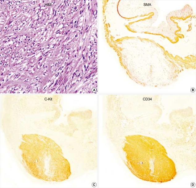
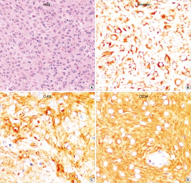
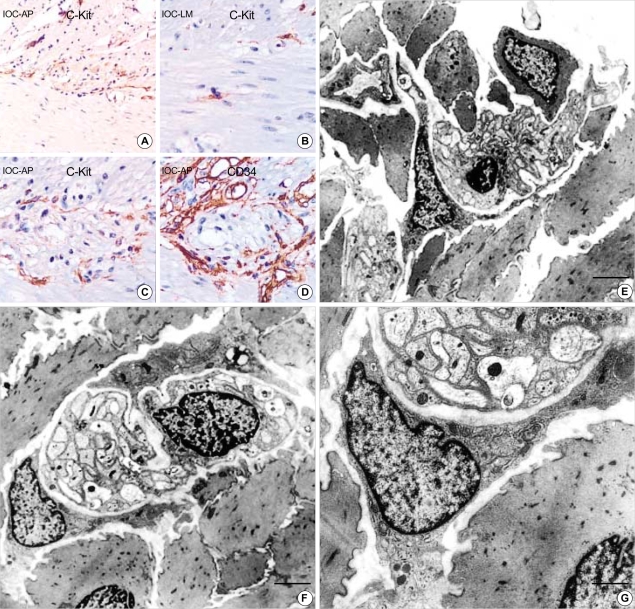
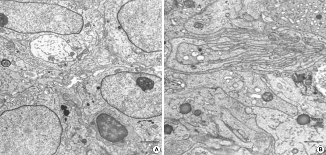
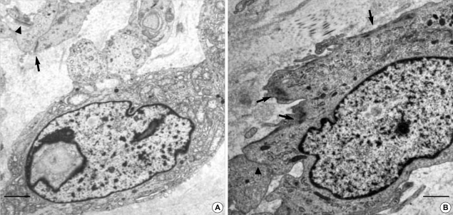
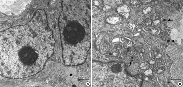
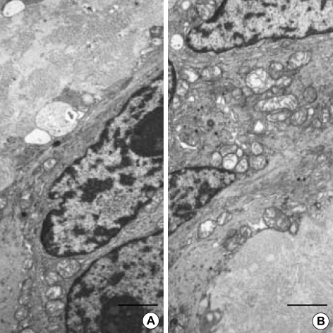
Gastrointestinal stromal tumors (GISTs) are the most common mesenchymal tumors in the gastrointestinal tract (GIT). Although interstitial cells of Cajal has been suggested as origin of this tumor, the cytological and ultrastructural features of GISTs are heterogeneous and unclear. A total 10 cases of normal gastrointestinal tissue (control), 13 GISTs of the stomach (8), small intestine (3), mesocolon (1) and liver (1), and 2 gastrointestinal autonomic nervous tumor (GANT) of small intestine were ultrastructurally studied. Normal interstitial cells of Cajal (ICC) were abundantly present around the myenteric plexuses or individually scattered through the wall of GIT. ICC was characterized by slender cytoplasmic processes, well-developed endoplasmic reticulum (ER), mitochondria, Golgi apparatus, caveolae and intermediate filaments. The GISTs and GANTs had overlapping ultrastructures. The most common and important ultrastructural features of GISTs were rich villous cytoplasmic processes, dispersed intermediate filaments and abundant SER, and those of GANTs were neurosecretory granules and skenoid fibers. Compared with ICC, the GISTs and GANTs had remarkably reduced caveolae and gap junctions. Our study suggested that ultrastructural analysis gives much information to investigate lineage differentiation of neoplastic cells and make a differential diagnosis of these tumors from other mesenchymal tumors and between GISTs and GANTs.
Figures

References
-
- Chan JK. Mesenchymal tumors of the gastrointestinal tract: a paradise for acronyms (STUMP, GIST, GANT, and now GIPACT), implication of c-kit in genesis, and yet another of the many emerging roles of the interstitial cell of Cajal in the pathogenesis of gastrointestinal diseases? Adv Anat Pathol. 1999;6:19–40. - PubMed
-
- Eyden B, Chorneyko KA, Shanks JH, Menasce LP, Banerjee SS. Contribution of electron microscopy to understanding cellular differentiation in mesenchymal tumors of the gastrointestinal tract: a study of 82 tumors. Ultrastruct Pathol. 2002;26:269–285. - PubMed
-
- Yantiss RK, Rosenberg AE, Selig MK, Nielsen GP. Gastrointestinal stromal tumors: an ultrastructural study. Int J Surg Pathol. 2002;10:101–113. - PubMed
-
- Liu P, Na J, Wang Y, He Q, Zhang Y, Tang X, Zou W. Study of gastrointestinal stromal tumors by light microscopy, electron microscopy and immunohistochemistry. Zhonghua Bing Li Xue Za Zhi. 2002;31:199–203. - PubMed
Publication types
MeSH terms
Substances
LinkOut - more resources
Full Text Sources
Miscellaneous
