. 2004 May;239(5):637-45; discussion 645-7.
doi: 10.1097/01.sla.0000124383.98416.8d.

Clinical genetic testing and early surgical intervention in patients with multiple endocrine neoplasia type 1 (MEN 1)

Affiliations

Clinical genetic testing and early surgical intervention in patients with multiple endocrine neoplasia type 1 (MEN 1)

Terry C Lairmore et al. Ann Surg. 2004 May.

Abstract

Objective: We sought to develop a comprehensive program for clinical genetic testing in a large group of extended families with multiple endocrine neoplasia type 1 (MEN 1), with the ultimate aim of early tumor detection and surgical intervention.

Summary background data: Germline mutations in the MEN1 tumor suppressor gene are responsible for the MEN 1 syndrome. Direct genetic testing for a disease-associated MEN1 mutation is now possible in selected families. The neuroendocrine tumors of the pancreas/duodenum and the intrathoracic neuroendocrine tumors that occur in MEN 1 carry a malignant potential. Importantly, these tumors arise in otherwise young healthy patients and are complicated by the potential for multifocality and involvement of multiple target tissues. The optimal screening methods and indications for early surgical intervention in genetically positive patients have yet to be defined.

Methods: Nine MEN 1 kindreds were included in the study. The mutations for each kindred were initially identified in the research laboratory. Subsequently, mutation detection was independently validated in the clinical Molecular Diagnostic Laboratory. Each patient in the study underwent formal genetic counseling before testing.

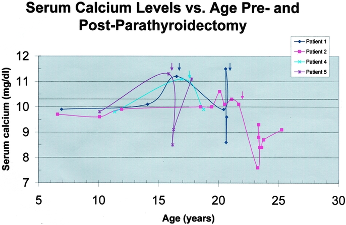
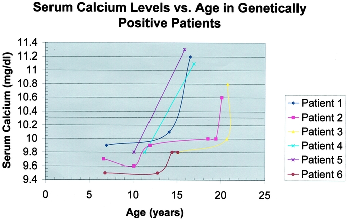
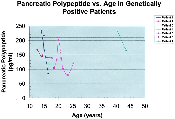
Results: Genetic testing was performed in 56 at-risk patients. Patients were stratified according to risk: Group I (n = 25), 50% risk, younger than 30 years old; Group II (n = 20), 50% risk, 30 years old or older; Group III (n = 11) 25% risk. Seven patients (age, 12 to 42 years; mean, 20.6 +/- 3.8 years) had a positive genetic test. Patients with a novel positive genetic test were in either Group I (n = 6) or Group II (n = 1) and have been followed for 35.8 +/- 2.0 months. Of the 7 genetically positive patients, hypercalcemia was either present at the time of diagnosis or developed during the period of follow-up in 6 patients. Four patients have undergone parathyroidectomy as early as age 16 years. One genetically positive patient has not yet developed hyperparathyroidism. Intensive biochemical screening in this select group of patients identified an elevated pancreatic polypeptide level and pancreatic tail mass lesion in a 15-year-old male who is asymptomatic and currently normocalcemic.

Conclusions: Genetic testing identifies patients harboring an MEN1 mutation before the development of clinical signs or symptoms of endocrine disease. When genetically positive patients are carefully studied prospectively, biochemical evidence of neoplasia can be detected an average of 10 years before clinically evident disease, allowing for early surgical intervention. Genetically positive individuals should undergo focused cancer surveillance for early detection of the potentially malignant neuroendocrine tumors that account for most of the disease-related morbidity and mortality.


Figures

None
FIGURE 1. The distribution of germline mutations along the MEN1 gene sequence in the study kindreds is shown. The MEN1 gene spans approximately 10 kb of genomic DNA and is made up of 10 exons (open rectangles). Exon 1 and the 3′ end of exon 10 are untranslated (shaded rectangles). The deletion, insertion, and nonsense mutations are depicted below the figure. The nomenclature is consistent with the recommendations from the Nomenclature Working Group (Antonarakis et al, 1998). The insertion and deletion mutations are numbered according to the nucleotide at which they occur relative to the A of the open reading frame. The nonsense mutations are numbered according to the codon in which they occur relative to the start methionine residue. The underlining denotes mutations that occur in more than 1 family. The specific mutations are further described in Table 2. Kindred 013 has an approximately 7-kb microdeletion of 1 allelic copy of the MEN1 gene encompassing the upstream 5′ region, exon 1, and most of exon 2 (data not shown) as depicted above the figure. (Modified from Hum Mutat 1999;13:175–185)
None
FIGURE 2. Serum calcium levels versus age in the 4 genetically positive patients undergoing parathyroidectomy are depicted graphically. Each patient's curve is represented by a different color and data point symbol according to the legend in the upper right corner. Serum calcium level (mg/dL) is plotted as a function of age (years). The upper limit of normal for calcium is indicated by the dotted line. The colored arrows indicate the time of parathyroidectomy for each patient. Patient 1 had identification of 3 glands in the neck at initial exploration and subsequently had successful removal of a deep mediastinal parathyroid gland at a second operation. Patient 5 has recurrent hypercalcemia following a 3.5-gland subtotal parathyroidectomy and an initial period of normocalcemia. All other patients are normocalcemic off all calcium and vitamin D supplementation at the final end point.
None
FIGURE 3. Serum calcium levels versus age in 6 of the 7 the genetically positive patients. Patient 7 has data only from ages 40 and 43 years. This patient is hypercalcemic but currently declines parathyroidectomy. Each patient's curve is represented by a different color and data point symbol according to the legend in the upper right corner. Serum calcium level (mg/dL) is plotted as a function of age (years). The upper limit of normal for calcium is indicated by the dotted line. In this selected subset of genetically positive patients followed prospectively, a rapid rise in calcium levels is evident between the ages of 10 and 15 years.
None
FIGURE 4. Age of onset of endocrine tumor expression in multiple endocrine neoplasia type 1 (MEN 1). Data derived from retrospective analysis for each endocrine organ hyperfunction in 130 cases of MEN1. Age at onset is the age at first symptom or, with tumors not causing symptoms, age at the time of the first abnormal finding on a screening test. The rate of diagnosis of hyperparathyroidism increased sharply between ages 16 and 20 years. (Reprinted with permission from Ann Intern Med 1998;129:484–494)
None
FIGURE 5. Serum pancreatic polypeptide levels versus age in the 7 genetically positive patients. Each patient's curve is represented by a different color and data point symbol according to the legend in the upper right corner. The pancreatic polypeptide level (pg/ml) is plotted as a function of age (years). The upper limit of normal for pancreatic polypeptide is dependent upon age and sex, but an average value for the age range plotted is indicated by the dotted line. Pancreatic polypeptide levels fluctuated significantly over time. No clear pattern or trend of progressive elevation was noted in this small subset of patients. Patient 6 had an elevated pancreatic polypeptide level detected at age 15 years. Computed tomography (CT) scanning in this patient reveals a probably neuroendocrine tumor of the pancreatic tail.

References

    1. Marx S, Spiegel AM, Skarulis MC, et al. Multiple endocrine neoplasia type 1: Clinical and genetic topics. Ann Intern Med. 1998;129:484–494. - PubMed
    1. Ballard HS, Frame B, Hartsock RJ. Familial multiple endocrine adenoma-peptic ulcer complex. Medicine. 1964;43:481–516. - PubMed
    1. Wilkinson S, Teh BT, Davey KR, et al. Cause of death in multiple endocrine neoplasia type 1. Arch Surg. 1993;128:683. - PubMed
    1. Doherty GM, Olson JA, Frisella MM, et al. Lethality of multiple endocrine neoplasia type 1. World J Surg. 1997;22:581–586. - PubMed
    1. Chandrasekharappa SC, Guru SC, Manickamp P, et al. Positional cloning of the gene for multiple endocrine neoplasia-type 1. Science. 1997;276:404–407. - PubMed

Publication types

LinkOut - more resources