

Correlation of amplification and overexpression of the c-myc oncogene in high-grade breast cancer: FISH, in situ hybridisation and immunohistochemical analyses
- PMID: 15083194
- PMCID: PMC2409705
- DOI: 10.1038/sj.bjc.6601703
Correlation of amplification and overexpression of the c-myc oncogene in high-grade breast cancer: FISH, in situ hybridisation and immunohistochemical analyses
Abstract
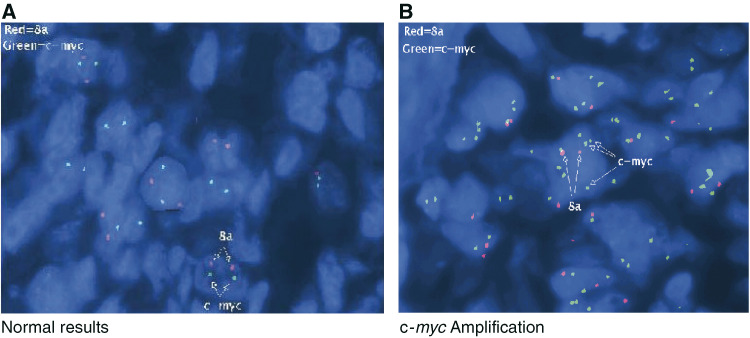
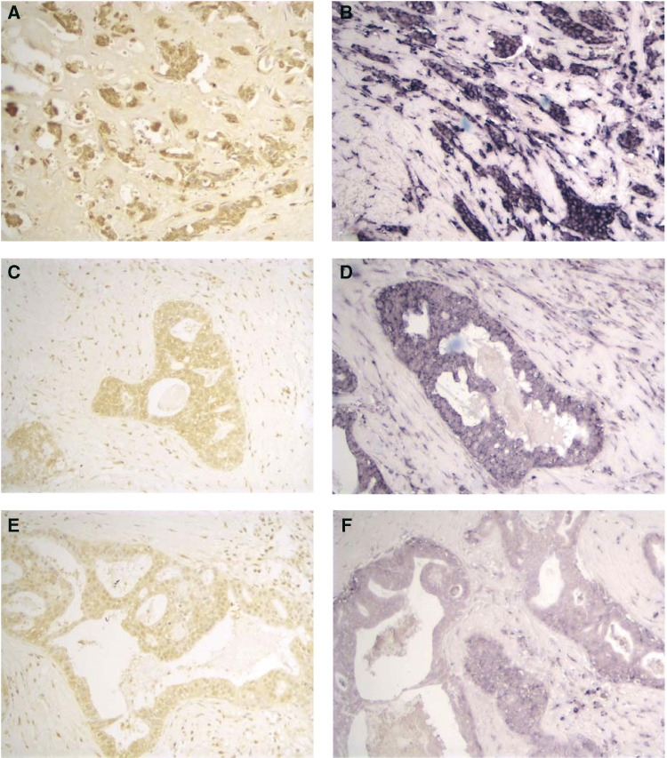
In this study, we analysed gene amplification, RNA expression and protein expression of the c-myc gene on archival tissue specimens of high-grade human breast cancer, using fluorescent in situ hybridisation (FISH), nonradioactive in situ hybridisation and immunohistochemistry. The specific question that we addressed was whether expression of c-Myc mRNA and protein were correlated with its gene copy amplification, as determined by FISH. Although c-Myc is one of the most commonly amplified oncogenes in human breast cancer, few studies have utilised in situ approaches to directly analyse the gene copy amplification, RNA transcription and protein expression on human breast tumour tissue sections. We now report that by using the sensitive FISH technique, a high proportion (70%) of high-grade breast carcinoma were amplified for the c-myc gene, irrespective of status of the oestrogen receptor. However, the level of amplification was low, ranging between one and four copies of gene gains, and the majority (84%) of the cases with this gene amplification gained only one to two copies. Approximately 92% of the cases were positive for c-myc RNA transcription, and essentially all demonstrated c-myc protein expression. In fact, a wide range of expression levels were detected. Statistically significant correlations were identified among the gene amplification indices, the RNA expression scores and protein expression scores. c-myc gene amplification, as detected by FISH, was significantly associated with expression of its mRNA, as measured by the intensity of in situ hybridisation in invasive cells (P=0.0067), and by the percentage of invasive cells positive for mRNA expression (P=0.0006). c-myc gene amplification was also correlated with the percentage of tumour cells which expressed high levels of its protein, as detected by immunohistochemistry in invasive cells (P=0.0016). Thus, although multiple mechanisms are known to regulate normal and aberrent expression of c-myc, in this study, where in situ methodologies were used to evaluate high-grade human breast cancers, gene amplification of c-myc appears to play a key role in regulating expression of its mRNA and protein.
Figures
References
-
- Augenlich L, Wadler S, Gorner G, Richards C, Ryan L, Multani A, Pathak S, Benson A, Haller D, Heerdt B (1997) Low-level c-myc amplification in human colonic carcinoma cell lines and tumors: a frequent, p53-independent mutation associated with improved outcome in a randomized multi-institutional trial. Cancer Res 57: 1769–1775 - PubMed
-
- Aulmann S, Bentz M, Sinn HP (2002) C-myc oncogene amplification in ductal carcinoma in situ of the breast. Breast Cancer Res Treat 74: 25–31 - PubMed
-
- Berns E, Klijn J, Van Putten W, van Staveren I, Portengen H, Foekens J (1992) c-myc amplification is a better prognostic factor than HER2/neu amplification in primary breast cancer. Cancer Res 52: 1107–1113 - PubMed
-
- Bieche I, Laurendeau I, Tozlu S, Olivi M, Vidaud D, Lidereau R, Vidaud M (1999) Quantitation of MYC gene expression in sporadic breast tumors with a real-time transcription-PCR assay. Cancer Res 59: 2759–2765 - PubMed
-
- Chana J, Cree I, Foss A, Hungerford J, Wilson G (1998a) The prognositc significance of c-myc oncogene expression in uveal melanoma. Melanoma Res 8: 139–144 - PubMed
Publication types
MeSH terms
Substances
Grants and funding
LinkOut - more resources
Full Text Sources
Other Literature Sources
Medical
