. 2004 Jul 15;381(Pt 2):547-59.
doi: 10.1042/BJ20040253.

Molecular analysis of muskelin identifies a conserved discoidin-like domain that contributes to protein self-association

Affiliations

Molecular analysis of muskelin identifies a conserved discoidin-like domain that contributes to protein self-association

Soren Prag et al. Biochem J. .

Abstract

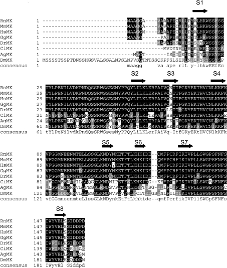
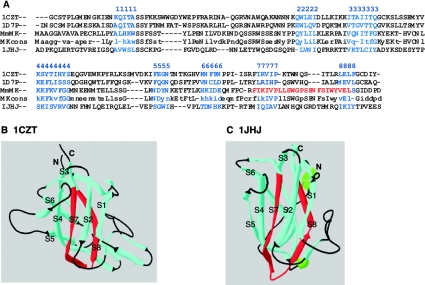
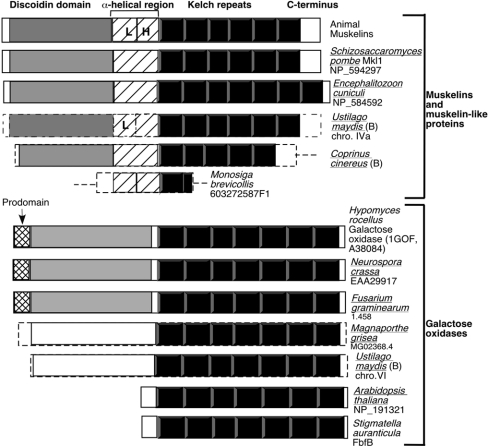
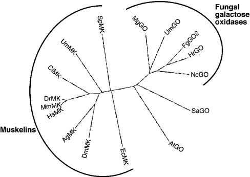
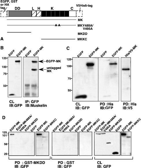
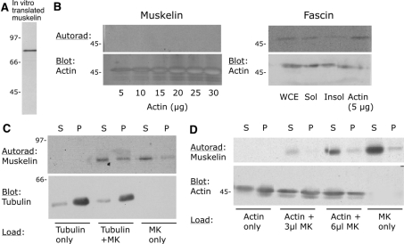
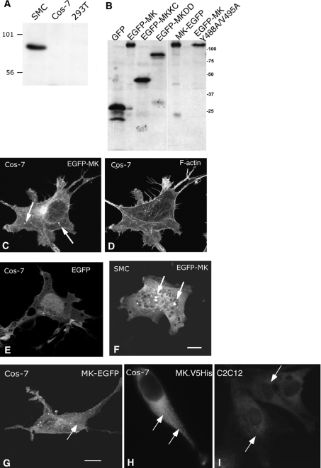
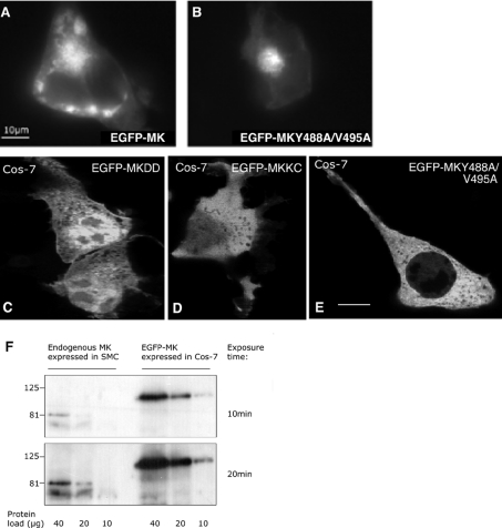
Muskelin is an intracellular protein with a C-terminal kelch-repeat domain that was initially characterized as having functional involvement in cell spreading on the extracellular matrix glycoprotein thrombospondin-1. As one approach to understanding the functional properties of muskelin, we have combined bioinformatic and biochemical studies. Through analysis of a new dataset of eight animal muskelins, we showed that the N-terminal region of the polypeptide corresponds to a predicted discoidin-like domain. This domain architecture is conserved in fungal muskelins and reveals a structural parallel between the muskelins and certain extracellular fungal galactose oxidases, although the phylogeny of the two groups appears distinct. In view of the fact that a number of kelch-repeat proteins have been shown to self-associate, co-immunoprecipitation, protein pull-down assays and studies of cellular localization were carried out with wild-type, deletion mutant and point mutant muskelins to investigate the roles of the discoidin-like and kelch-repeat domains. We obtained evidence for cis- and trans-interactions between the two domains. These studies provide evidence that muskelin self-associates through a head-to-tail mechanism involving the discoidin-like domain.


Figures

Figure 1
Figure 1. CLUSTALW alignment of the N-terminal regions of eight animal muskelins
The N-terminal sequences of Anopheles gambiae (Ag; GenBank XP_314298 and BM606168), Ciona intestinalis (Ci; BW210776 and other ESTs in cluster 05014r1), Danio rerio (Dr; AF418017), Drosophila melanogaster (Dm; AF049333), Gallus gallus (Gg; AJ451691), Homo sapiens (Hs; NP_037387), Mus muscularis (Mm; NP_038819) and Rattus norvegicus (Rn; NM_031359) muskelins (MKs) were aligned in CLUSTALW and are presented in Boxshade. Black shading indicates identical residues, grey shading indicates conservative substitutions, and no shading indicates unrelated amino acids. The 50% identity consensus sequence is shown below the alignment. Blocks corresponding to the eight β-strands of a discoidin domain β-barrel, S1–S8, are indicated by arrows (see also Figure 2).
Figure 2
Figure 2. The muskelin N-terminal region is predicted to fold as a discoidin domain
(A) Alignment by CLUSTALW of the N-terminal region of mouse muskelin (MmMK) and the 50% identity consensus sequence (MKcons) with three discoidin domain structures from PDB. 1CZT, C2 domain of coagulation factor V; 1D7P, C2 domain of coagulation factor VIII; 1JHJ, anaphase-promoting complex subunit 10. The eight β-strands of the β-barrel (numbered here 1–8) were assigned from the CDD and by inspection of the primary sequences against the structures in PDB, and are marked in blue. The MIND motif in muskelin is marked in red. (B, C) Ribbon diagrams of the discoidin domains of 1CZT (B) and 1JHJ (C). In each structure, β-strands are in blue (with the strands of the β-barrel labelled S1–S8), loops are in black, and the regions aligned with the muskelin MIND motif (as shown in A) are in red.
Figure 3
Figure 3. Domain architecture of muskelins and extracellular galactose oxidases
In each stick model, the predicted discoidin-like domain is shown in grey, the kelch repeats in black, the central α-helical region of muskelin with diagonal lines, the prodomain of galactose oxidase as cross-hatching, and unrelated portions of the polypeptides in white. L, LisH motif; H, CTLH motif (as identified in SMART). The broken lines for Ustilago maydis, Coprinus cinereus, Magnaporthe grisea and Monosiga brevicollis N- and C-terminal ends indicate that the ends of the ORFs are not definitively assigned. The seven kelch repeats in NP_191321 and AAL25195 were identified by multiple sequence alignment with A38084 and assignment of the kelch repeats according to the 1GOF structure. NP_191321 and AAL25195 contain predicted signal peptides. B, basidiomycote. Organisms for which the complete genome sequence is available are underlined.
Figure 4
Figure 4. Phylogenetic relationships of muskelins and galactose oxidases
Six kelch repeats from each of the indicated species of muskelin (MK) or galactose oxidase (GO) were selected for analysis as described in the Materials and methods section and aligned in CLUSTALW, and are presented as an unrooted dendrogram in DRAWTREE. The incomplete Coprinus cinereus muskelin sequence was not included. Key: Ag, Anopheles gambiae; At, Arabidopsis thaliana; Ci, Ciona intestinalis; Dr, Danio rerio; Dm, Drosophila melanogaster; Ec, Encephalitizoon cuniculi; Fg, Fusarium graminearum; Hr, Hypomyces rocellus; Hs, Homo sapiens; Mg, Magnaporthe grisea; Mm, Mus muscularis; Nc, Neurospora crassa; Sa, Stigmatella aurantiaca; Sp, Schizosaccharomyces pombe; Um, Ustilago maydis.
Figure 5
Figure 5. Biochemical analysis of muskelin self-association
(A) Schematic diagram of muskelin constructs, their designations and the various N- and C-terminal tags utilized. HA, haemagglutinin; DD, discoidin-like domain; L, LisH motif; H, CTLH motif; K, kelch repeats; C, C-terminal region. (B) Co-immunoprecipitation of muskelin with EGFP–MK. Lysates from 293T cells expressing EGFP or EGFP–MK were mixed 1:1 (v/v) with SMC lysates and immunoprecipitated (IP) for GFP. Bound proteins were resolved on 10% (w/v) polyacrylamide gels, transferred to a PVDF membrane and probed with antibodies to muskelin. Expression of the transiently expressed proteins was monitored by immunoblot (IB) of cell lysates with anti-GFP antibody (CL). (C) Specific pull-down (PD) of EGFP–MK with MK–V5His6. Lysates of 293T cells transiently expressing MK–V5His6 were mixed 1:1 (v/v) with lysates from cells expressing EGFP or EGFP–MK and incubated with nickel beads. Bead-bound proteins were resolved on 10% (w/v) polyacrylamide gels, transferred to a PVDF membrane and probed with antibodies to polyhistidine or GFP. Expression of the transiently expressed proteins was monitored by immunoblotting (IB) of cell lysates (CL). (D) Mapping requirements for pull-down (PD) by GST–MKDD. Aliquots of 400 μg of detergent lysates of 293T cells transiently transfected with the indicated EGFP–MK proteins were incubated with either GST–MKDD or GST-loaded glutathione–agarose beads. Aliquots of 45 μg of the lysates were also taken to monitor protein levels (CL). The bound proteins were resolved on SDS/10%-polyacrylamide gels, transferred to a PVDF membrane and probed with antibody to GFP. Only wild-type EGFP–MK and EGFP–MKKC bound to GST–MKDD, and there was no binding to GST. The results are representative of three independent experiments.
Figure 6
Figure 6. Muskelin does not bind actin or tubulin
(A) Analysis by SDS/PAGE and autoradiography of [35S]methionine-labelled in vitro-translated muskelin, as used for the experiments shown in (B)–(D). (B) Analysis of the binding of muskelin or fascin to G-actin, detected by blot overlay. In the left-hand panel, the indicated concentrations of actin were resolved on an SDS/12.5%-PAGE gel, transferred to nitrocellulose, renatured and incubated with radiolabelled muskelin. In the right-hand panel, the same procedure was followed using 50 μg of whole-cell extract (WCE), or 50 μg of detergent-soluble (Sol) or -insoluble (Insol) fractions, or 5 μg of G-actin, and the blot was incubated with radiolabelled in vitro-translated fascin. (C, D) Results of co-sedimentation assays carried out as described in the Materials and methods section for microtubule assembly (C) or F-actin polymerization (D). Equal aliquots of supernatant (S) or pellet (P) were resolved on SDS/12.5%-polyacrylamide gels, transferred to nitrocellulose and probed with antibodies to tubulin or actin as appropriate. Muskelin was detected by autoradiographic exposure of the same blots (Autorad). The results are representative of three independent experiments. In all panels, molecular mass markers are in kDa.
Figure 7
Figure 7. Expression and localization of muskelin in cells
(A) Western blot of an SDS/10%-polyacrylamide gel loaded with 40 μg of 1% Triton X-100 cell extracts from SMCs, Cos-7 cells and 293T cells, probed with anti-muskelin antibodies. (B) Immunoblot showing relative expression of EGFP–MK proteins, detected by anti-GFP antibody. Protein designations are as in (A). Molecular mass markers are in kDa. (C)–(I) Confocal images of the distribution of transiently expressed EGFP–MK (C, D), MK–EGFP (G) or MK–V5His6 (H) in Cos-7 cells, or EGFP–MK in SMCs (F). Panels (C) and (D) show a Cos-7 cell co-stained for F-actin. (E) Distribution of EGFP in Cos-7 cells. (I) Endogenous muskelin of C2C12 cells (as XY section from Z stack). Arrows in panels (C) and (F)–(I) indicate the muskelin particles of various sizes observed in the cells. Bars=10 μm.
Figure 8
Figure 8. Roles of the discoidin-like domain and the β-propeller in particle formation in cells
(A, B) Cos-7 cells expressing EGFP–MK (A) or EGFP–MK488A/V495A (B) were treated with 100 μM PSI for 12 h before fixation. Large muskelin aggregates were formed in the cells. (C)–(E) Localization of muskelin mutants. Confocal XY sections of Cos-7 cells transiently expressing EGFP–MKDD (C), EGFP–MKKC (D) or EGFP–MK-Y488A/V495A (E) are shown. None of these proteins formed particles. Bars=10 μm. (F) Expression of EGFP–MK in Cos-7 cells relative to endogenous muskelin in SMCs. The indicated protein loads of whole-cell extracts were resolved on an SDS/10%-polyacrylamide gel, transferred to a PVDF membrane and probed with anti-muskelin antibodies. Two ECL® exposure times were taken for quantification of expression using Scion Image (NIH). Pixel intensity per area was quantified at each exposure time (EGFP–MK signal from a 10 μg protein load compared with muskelin signal from 40 and 20 μg protein loads at 20 min exposure time) and mean values calculated.

Similar articles

Cited by

References

    1. Adams J. C., Seed B., Lawler J. Muskelin, a novel intracellular mediator of cell adhesive and cytoskeletal responses to thrombospondin-1. EMBO J. 1998;17:4964–4974. - PMC - PubMed
    1. Adams J. C., Zhang L. cDNA cloning of human muskelin and localization of the muskelin (MKLN1) gene to human chromosome 7q32 and mouse chromosome 6 B1/B2 by physical mapping and FISH. Cytogenet. Cell Genet. 1999;87:19–21. - PubMed
    1. Adams J. Characterization of a Drosophila melanogaster orthologue of muskelin. Gene. 2002;297:69–78. - PubMed
    1. Cahana A., Escamez T., Nowakowski R. S., Hayes N. L., Giacobini M., von Holst A., Shmueli O., Sapir T., McConnell S. K., Wurst W., et al. Targeted mutagenesis of Lis1 disrupts cortical development and LIS1 homodimerization. Proc. Natl. Acad. Sci. U.S.A. 2001;98:6429–6434. - PMC - PubMed
    1. Smith D. S., Niethammer M., Ayala R., Zhou Y., Gambello M. J., Wynshaw-Boris A., Tsai L. H. Regulation of cytoplasmic dynein behaviour and microtubule organization by mammalian Lis1. Nat. Cell Biol. 2000;2:767–775. - PubMed

Publication types

MeSH terms

Associated data