

Review
doi: 10.1111/j.1542-474X.2004.92538.x.
Evolving myocardial infarction with ST elevation: ups and downs of ST in different leads identifies the culprit artery and location of the occlusion
Affiliations
- PMID: 15084217
- PMCID: PMC6931954
- DOI: 10.1111/j.1542-474X.2004.92538.x
Item in Clipboard
Review
Evolving myocardial infarction with ST elevation: ups and downs of ST in different leads identifies the culprit artery and location of the occlusion
Ann Noninvasive Electrocardiol.
2004 Apr.
No abstract available
Figures
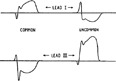
Predicting artery occlusion from ECG changes (taken from Lewis
9
).
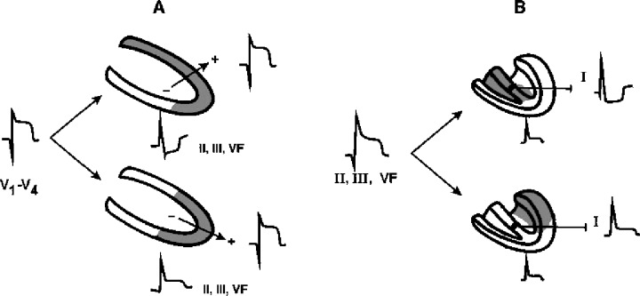
(A) Shows how in the case of ST elevation in precordial leads as a consequence of occlusion in LAD, the ST changes in reciprocal leads (II, III, VF) allow to identify whether the occlusion is in proximal (above) or distal LAD (below), and (B) shows how in the case of ST elevation in II, III, and VF the changes of ST in other leads (lead I) give us information on if the inferoposterior wall infarct is due to RCA (above) or LCX (below) occlusion.
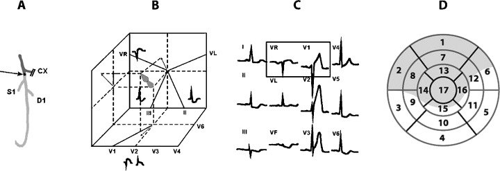
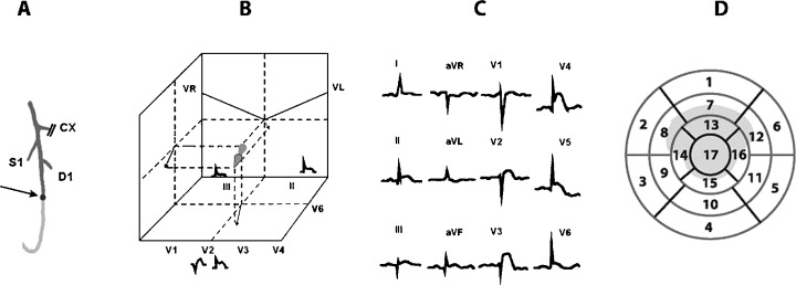
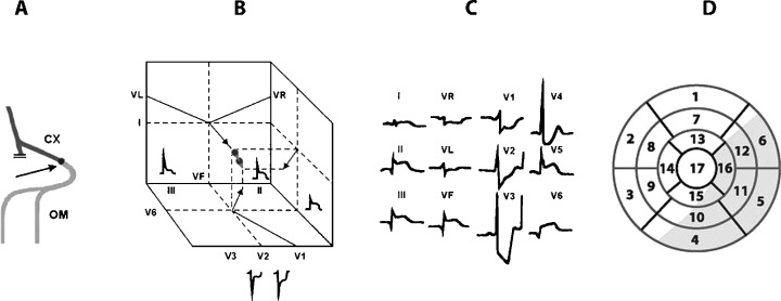
The location of the occlusion (A), vectors of injury (B), ECG changes (C), and segments affected in eye bull view (D), in case of occlusion of LAD proximal to D1 and S1.
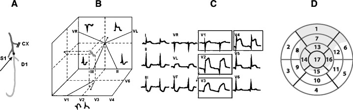
The location of the occlusion (A), vectors of injury (B), ECG changes (C), and segments affected in eye bull view (D) in case of occlusion LAD proximal to D1 but distal to S1.
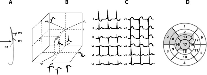
The location of the occlusion (A), vectors of injury (B), ECG changes (C), and segments affected in eye bull view (D) in case of occlusion LAD distal to S1 and D1.
The location of the occlusion (A), vectors of injury (B), ECG changes (C), and segments affected in eye bull view (D) in case of occlusion LAD distal to D1 but proximal to S1.
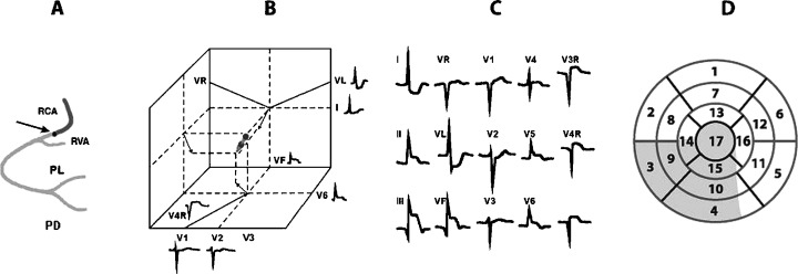
The location of the occlusion (A), vectors of injury (B), ECG changes (C), and segments affected in eye bull view (D) in case of proximal occlusion of RCA, before the artery of the right ventricle.
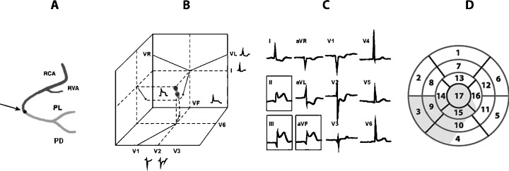
The location of the occlusion (A), vectors of injury (B), ECG changes (C), and segments affected in eye bull view (D) in case of occlusion of RCA distal to the artery of right ventricle.
The location of the occlusion (A), vectors of injury (B), ECG changes (C), and segments affected in eye bull view (D) in case of occlusion of Cx.
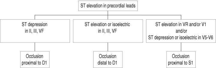
The algorithm to follow for prediction of the place of occlusion in LAD artery in case of evolving MI with ST elevation in precordial leads (see text).
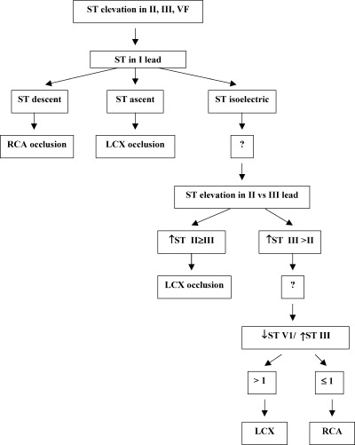
The algorithm to follow for prediction of the culprit artery (RCA vs. LCX) in case of evolving MI with ST elevation in inferior leads (see text).
References
-
- Myers GB, Klein HA, Stofer BE. Correlation of electrocardiographic findings in anteroseptal infarction. Am Heart J 1948;36: 535–575. - PubMed
-
- Startt Selvester RH, Wagner GS, Ideker RE. Myocardial infarction In McFarlane PW, Veitch Lawrie TD. (eds.): Comprehensive Electrocardiology. New York , Pergamon Press, 1989.
-
- Chou T. Electrocardiography in Clinical Practice. New York , Grune&Stratton, 1979.
-
- Wagner G. Marriott's Practical Electrocardiography. Philadelphia , Lippincott Williams and Wilkins, 2001.
-
- Sclarowsky S. Electrocardiography of Acute Myocardial Ischaemia. London , Martin Dunitz , 1999.
Publication types
MeSH terms
LinkOut - more resources
Full Text Sources
Medical
