

DAX-1 expression in human breast cancer: comparison with estrogen receptors ER-alpha, ER-beta and androgen receptor status
- PMID: 15084237
- PMCID: PMC400665
- DOI: 10.1186/bcr766
DAX-1 expression in human breast cancer: comparison with estrogen receptors ER-alpha, ER-beta and androgen receptor status
Abstract
Background: So far there have been no reports on the expression pattern of DAX-1 (dosage-sensitive sex reversal, adrenal hypoplasia critical region, on chromosome X, gene 1) in human breast cells and its relationship to the estrogen receptors, ER-alpha and ER-beta, and the androgen receptor (AR).
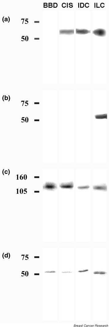
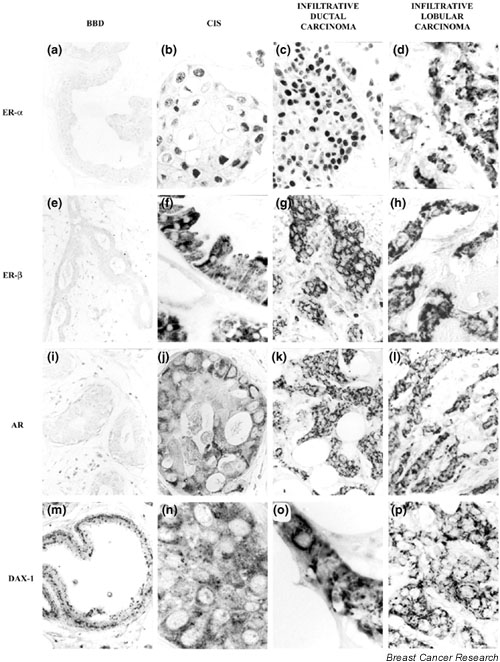
Methods: In this study we evaluated, by immunohistochemistry and Western blot analysis, the presence and distribution of DAX-1 in benign breast disease (BBD), in situ carcinoma (CIS), and ductal and lobular breast carcinomas.
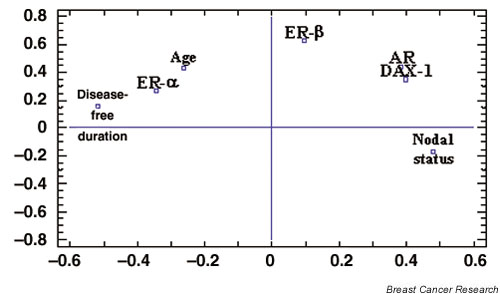
Results: In BBD and breast carcinomas, DAX-1 was present in both the nuclei and the cytoplasm of epithelial cells, although in infiltrative carcinomas the percentage of nuclear immunoreaction was higher than in CIS. An important relation was observed between DAX-1 and AR expression and between this orphan receptor and nodal status.
Conclusion: DAX-1 might modify the AR and ER-beta intracellular location, and because a direct positive relation between the expression of these three receptors was found it could be assumed that the presence of DAX-1 in neoplastic cells might indicate a possible failure of endocrine therapies.
Figures
Similar articles
-
[Expression of oestrogen receptor-beta in invasive breast tumours].Magy Onkol. 2004;48(1):63-9. Epub 2004 Apr 23. Magy Onkol. 2004. PMID: 15105898 Hungarian.
-
[Expression and intranuclear distribution of nucleolin in estrogen receptor-negative and estrogen receptor-positive breast cancers in women measured by laser scanning cytometry].Ann Acad Med Stetin. 2006;52(2):23-32. Ann Acad Med Stetin. 2006. PMID: 17633394 Polish.
-
Distribution, frequency, and quantitative analysis of estrogen, progesterone, androgen, and glucocorticoid receptors in human breast cancer.Cancer Res. 1979 May;39(5):1447-54. Cancer Res. 1979. PMID: 427788
-
The gene responsible for adrenal hypoplasia congenita, DAX-1, encodes a nuclear hormone receptor that defines a new class within the superfamily.Recent Prog Horm Res. 1996;51:241-59; discussion 259-60. Recent Prog Horm Res. 1996. PMID: 8701082 Review.
-
Molecular mechanisms of DAX1 action.Mol Genet Metab. 2004 Sep-Oct;83(1-2):60-73. doi: 10.1016/j.ymgme.2004.07.018. Mol Genet Metab. 2004. PMID: 15464421 Review.
Cited by
-
Expression of AIB1 protein as a prognostic factor in breast cancer.World J Surg Oncol. 2011 Oct 29;9:139. doi: 10.1186/1477-7819-9-139. World J Surg Oncol. 2011. PMID: 22035181 Free PMC article.
-
Orphan nuclear receptors in breast cancer pathogenesis and therapeutic response.Endocr Relat Cancer. 2010 Aug 16;17(3):R213-31. doi: 10.1677/ERC-10-0058. Print 2010 Sep. Endocr Relat Cancer. 2010. PMID: 20576803 Free PMC article. Review.
-
Bioanalytical LC-MS Method for the Quantification of Plasma Androgens and Androgen Glucuronides in Breast Cancer.J Chromatogr Sci. 2016 Apr;54(4):583-92. doi: 10.1093/chromsci/bmv190. Epub 2016 Jan 12. J Chromatogr Sci. 2016. PMID: 26762957 Free PMC article.
-
Hormonal Homologies between Canine Mammary Cancer and Human Breast Cancer in a Series of Cases.Vet Sci. 2022 Jul 29;9(8):395. doi: 10.3390/vetsci9080395. Vet Sci. 2022. PMID: 36006309 Free PMC article.
-
Minireview: role of orphan nuclear receptors in cancer and potential as drug targets.Mol Endocrinol. 2014 Feb;28(2):157-72. doi: 10.1210/me.2013-1291. Epub 2013 Dec 2. Mol Endocrinol. 2014. PMID: 24295738 Free PMC article. Review.
References
-
- Palmieri C, Cheng GJ, Saji S, Zelada-Hedman M, Wärri A, Weihua Z, Van Noorden S, Wahlstrom T, Coombes RC, Warner M, Gustafsson JA. Estrogen receptor beta in breast cancer. Endocrine-Related Cancer. 2002;9:1–13. - PubMed
-
- Burak WE, Jr, Quinn AL, Farrar WB, Brueggeneier RW. Androgens influence estrogen-induced responses in human breast carcinoma cells through cytochrome P450 aromatase. Breast Cancer Res Treat. 1997;44:57–64. - PubMed
-
- McKenna NJ, Lanz RB, O'Malley BW. Nuclear receptor coregulators: cellular and molecular biology. Endocr Rev. 1999;20:321–344. - PubMed
Publication types
MeSH terms
Substances
LinkOut - more resources
Full Text Sources
Medical
Research Materials
