Isolated left ventricular apical hypoplasia: a new congenital anomaly described with cardiac tomography
- PMID: 15084556
- PMCID: PMC1768218
- DOI: 10.1136/hrt.2003.010637
Isolated left ventricular apical hypoplasia: a new congenital anomaly described with cardiac tomography
Abstract
Objective: To describe cardiac tomography findings of an apparently new, presumably congenital, left ventricular (LV) abnormality noted consistently in three patients.
Patients: Three patients presenting with non-specific symptoms including fatigue, shortness of breath, or chest discomfort were evaluated with cardiac tomography for cardiac structure and function.
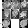
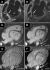
Results: Findings from the three patients were very similar: a truncated and spherical LV with abnormal diastolic and systolic function, invagination of fatty material into the myocardium of the defective LV apex, origin of a complex papillary network in the anteroapical LV, and an elongated right ventricle wrapping around the deficient apex.
Conclusions: Isolated LV apical hypoplasia is a unique, presumably congenital, cardiac anomaly that is an important condition to recognise.
Figures


References
-
- Hirsch R, Kilner PJ, Connelly MS, et al. Diagnosis in adolescents and adults with congenital heart disease: prospective assessment of individual and combined roles of magnetic resonance imaging and transesophageal echocardiography. Circulation 1994;90:2937–51. - PubMed
-
- Sardanelli F, Molinari G, Petillo A, et al. MRI in hypertrophic cardiomyopathy: a morphofunctional study. J Comput Assist Tomogr 1993;17:862–72. - PubMed
-
- Masui T, Finck S, Higgins CB. Constrictive pericarditis and restrictive cardiomyopathy: evaluation with MR imaging. Radiology 1992;182:369–73. - PubMed
-
- Doherty NE 3rd, Fujita N, Caputo GR, et al. easurement of right ventricular mass in normal and dilated cardiomyopathic ventricles using cine magnetic resonance imaging. Am J Cardiol 1992;69:1223–8. - PubMed
Publication types
MeSH terms
LinkOut - more resources
Full Text Sources
Miscellaneous
