. 2004 Apr 14;24(15):3801-9.
doi: 10.1523/JNEUROSCI.5543-03.2004.

Dimeric amyloid beta protein rapidly accumulates in lipid rafts followed by apolipoprotein E and phosphorylated tau accumulation in the Tg2576 mouse model of Alzheimer's disease

Affiliations

Dimeric amyloid beta protein rapidly accumulates in lipid rafts followed by apolipoprotein E and phosphorylated tau accumulation in the Tg2576 mouse model of Alzheimer's disease

Takeshi Kawarabayashi et al. J Neurosci. .

Abstract

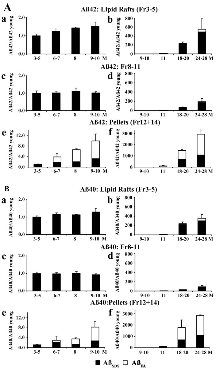
To investigate lipid rafts as a site where amyloid beta protein (Abeta) oligomers might accumulate and cause toxicity in Alzheimer's disease (AD), we analyzed Abeta in the Tg2576 transgenic mouse model of AD. Abeta was highly concentrated in lipid rafts, which comprise a small fraction of brain volume but contain 27% of brain Abeta42 and 24% of Abeta40 in young mice. In the Tg2576 model, memory impairment begins at 6 months before amyloid plaques are visible. Here we show that Abeta dimers appear in lipid rafts at 6 months and that raft Abeta, which is primarily dimeric, rapidly accumulates reaching levels >500x those in young mice by 24-28 months. A similar large accumulation of dimeric Abeta was observed in lipid rafts from AD brain. In contrast to extracellular amyloid fibrils, which are SDS-insoluble, virtually all Abeta in lipid rafts is SDS soluble. Coupled with recent studies showing that synthetic and naturally occurring Abeta oligomers can inhibit hippocampal long-term potentiation, the in vivo age-dependent accumulation of SDS-soluble Abeta dimers in lipid rafts at the time when memory impairment begins in Tg2576 mice provides strong evidence linking Abeta oligomers to memory impairment. After dimeric Abeta began to accumulate in lipid rafts of the Tg2576 brain, apolipoprotein E (ApoE) and then phosphorylated tau accumulated. A similar increase in ApoE and a large increase in phosphorylated tau was observed in lipid rafts from AD brain. These findings suggest that lipid rafts may be an important site for interaction between dimeric Abeta, ApoE, and tau.


Figures

Figure 1.
Figure 1.
Characterization of lipid rafts from 3-month-old Tg2576 mouse brains. A, Western blotting of sucrose gradient centrifuge fractions. Lanes 1–11 show the 11 fractions from the sucrose gradient. The lipid raft markers, flotillin and GM1 ganglioside, localized in fraction 4. The ER marker, calnexin, localized in fractions 8–11. APP, CTF of APP, BACE1, PS1-N, PS1-C, neprilysin, and ApoE localized in lipid rafts as well as in fractions 8–11. B, Fraction 4 was composed of vesicular structures of various sizes by EM. Scale bar, 600 nm. C, D, Levels of Aβ42SDS (C) and Aβ40SDS (D) in fractions 1–11 from 3-month-old Tg2576 brains (n = 6). E, Immunoblotting of Aβ in lipid rafts from 3-month-old transgenic brains. Synthetic Aβ1–40 (a, 2,000 fmol; b, 500 fmol; c, 125 fmol), fraction 4 of 3-month-old transgenic brains (d, 0.5 ml; e, 2 ml; f, 2 ml) were immunoprecipitated with 3160 (anti-Aβ1–40). Proteins were separated on 16% tricine gels, transferred to polyvinylidene difluoride membrane, and detected with BAN-50 (anti-Aβ1–16, lanes a–e) or 4G8 (anti-Aβ17–24, lane f). The arrowhead identifies CTFβ, and the arrow identifies Aβ monomers.
Figure 2.
Figure 2.
Age-dependent changes in Aβ42 (A) and Aβ40 (B) in Tg2576 mouse brain. The figures on the left show brain Aβ in 3- to 10-month-old mice; those on the right show brain Aβ in 9- to 28-month-old mice. Fractions shown are lipid raft Aβ (fractions 3–5; a, b), Triton-soluble Aβ (fractions 8–11; c, d), and pellet Aβ (fractions 12 + 14; e, f). The Aβ measured in each fraction at each age is expressed in terms of the SDS-soluble Aβ (AβSDS) in that fraction at 3–5 months (Aβ in fraction at specified age/AβSDS in fraction at 3–5 months). SDS-soluble Aβ (AβSDS) is shown in filled columns; SDS-insoluble Aβ that was solubilized in formic acid (AβFA) is shown in open columns.
Figure 3.
Figure 3.
Electron microscopy of Aβ42, Aβ40, and Aβ fibrils in the lipid raft fraction and pellets from 28-month-old Tg2576 brain. a–c, IEM showed that vesicular profiles in fraction 4 were labeled with anti-Aβ antibodies (a, BAN-50; b, BA-27; c, BC-05). No labeling was observed in control experiments in which anti-Aβ antibodies were omitted (data not shown). d, Many amyloid fibrils stained with 3160 coupled with 5 nm gold particles were present in fraction 12 (high speed gradient pellet). e, Amyloid cores were detected in fraction 14 (low-speed Triton pellet). Scale bar: a–d, 150 nm; e, 300.
Figure 4.
Figure 4.
Aβ42 and Aβ40 in control, pathological aging (PA), and AD brains. The figures on the left show Aβ42, and those on the right Aβ40 in control (n = 2), pathologic aging (n = 2), and AD (n=6) brains. Fractions shown are lipid raft Aβ (fractions 3–5; A,D), Triton-soluble Aβ (fractions 8–11; B, E), and pellet Aβ (fractions 12 + 14; C, F). SDS-soluble Aβ (AβSDS) is shown in filled columns; SDS-insoluble Aβ that was solubilized in formic acid (AβFA) is shown in open columns. Standard error bars are not shown for pathologic aging and controls because only two brains were analyzed in each instance.
Figure 5.
Figure 5.
Aβ dimers in lipid rafts from Tg2576 and human brains. A, Fractions 1–11 from 11-month-old Tg2576 brain. Ba, Fraction 4 from 4-, 8-, 10-, and 12-month-old Tg2576 brains. Bb, Fraction 4 from 5-, 6-, 7-, 8-, 10-, and 11-month-old Tg2576 brains labeled with 4G8. The Aβ in A and B was immunoprecipitated with polyclonal antibody 3160 and then immunoblotted with 4G8, a monoclonal antibody that detects both monomers and dimers. Dimers were the predominant form of Aβ detected in lipid rafts. Aβ dimers were detected at 6 months and progressively accumulated thereafter. C, Epitope mapping of fraction 4 from 17-month-old Tg2576 brain using anti-Aβ and anti-APP antibodies. Aβ was immunoprecipitated with 3160 and then immunoblotted with Z31preA (B), BAN-50 (C), 4G8 (D), BA-27 (E), BC-05 (F), and O443 (G). Lane A is synthetic Aβ40 (100 pmol) immunoprecipitated with 3160 and detected by BAN-50. The 8 kDa Aβ dimer and the 4 kDa Aβ monomer were detected by all anti-Aβ antibodies. BA-27 specifically detects the C terminus of Aβ40 and BC-05 is selective for the C terminus of Aβ42, so the labeling by these antibodies shows that the 8 kDa protein terminates at Aβ40 or at Aβ42 on the carboxylside. Z31preA, which recognizes the APP epitope just amino to Aβ failed to detect the 8 kDa band, showing that the 8 kDa band is dimeric Aβ and not an Aβ-bearing APP fragment that extends past Aβ on the amino side. The 8 kDa Aβ dimers were also not detected by anti-C terminus of APP (O443). Because 3160 recognizes the N-terminal region of Aβ, p3 was not detected with this immunoprecipitation. D, Fractions 1–11 from AD, pathological aging (PA), and control brains. The Aβ in the fractions shown in D was directly immunoblotted with 4G8.
Figure 6.
Figure 6.
Accumulation of ApoE and phosphorylated tau in lipid rafts. A, One microliter of fraction 4 at 3, 4, 5, 8, 12, 18, 24, and 28 months was analyzed by immunoblot with antibodies to flotillin, APP, ApoE, WKS46 (aphosphorylation-independent antibody against tau 359–370), and PHF-1 (an antibody to tau phosphorylated at pSer396/pSer404) sites. The amount of flotillin and APP did not change with aging. ApoE increased substantially beginning at 12 months. Total tau detected by WKS46 did not change with aging, whereas phosphorylated-tau stained by PHF-1 increased beginning at 18 months. Sucrose gradient centrifuge fractions (1–11) from AD and control brains were analyzed by immunoblot using anti-flotillin antibody (B), anti-human ApoE antibody (C), or PHF-1 (D). Note the accumulation of ApoE in lipid rafts (fraction 4) from AD as compared with control brains and the intensely labeled smear of phosphorylated tau in lipid rafts from AD but not control brain.

References

    1. Avdulov NA, Chochina SV, Igbavboa U, Warden CS, Vassiliev AV, Wood WG (1997) Lipid binding to amyloid β-peptide aggregates: preferential binding of cholesterol as compared with phosphatidylcholine and fatty acids. J Neurochem 69: 1746–1752. - PubMed
    1. Bales KR, Verina T, Dodel RC, Du Y, Altstiel L, Bender M, Hyslop P, Johnstone EM, Little SP, Cummins DJ, Piccardo P, Ghetti B, Paul SM (1997) Lack of apolipoprotein E dramatically reduces amyloid β-peptide deposition. Nat Genet 17: 263–264. - PubMed
    1. Bickel PE, Scherer PE, Schnitzer JE, Oh P, Lisanti MP, Lodish HF (1997) Flotillin and epidermal surface antigen define a new family of caveolae-associated integral membrane proteins. J Biol Chem 272: 13793–13802. - PubMed
    1. Brandt R, Leger J, Lee G (1995) Interaction of tau with the neural plasma membrane mediated by tau's amino-terminal projection domain. J Cell Biol 131: 1327–1340. - PMC - PubMed
    1. Brown DA, London E (1998) Functions of lipid rafts in biological membranes. Annu Rev Cell Dev Biol 14: 111–136. - PubMed

Publication types

MeSH terms