. 2004 Apr 14;24(15):3837-49.
doi: 10.1523/JNEUROSCI.5539-03.2004.

Sequential release of GABA by exocytosis and reversed uptake leads to neuronal swelling in simulated ischemia of hippocampal slices

Affiliations

Sequential release of GABA by exocytosis and reversed uptake leads to neuronal swelling in simulated ischemia of hippocampal slices

Nicola J Allen et al. J Neurosci. .

Abstract

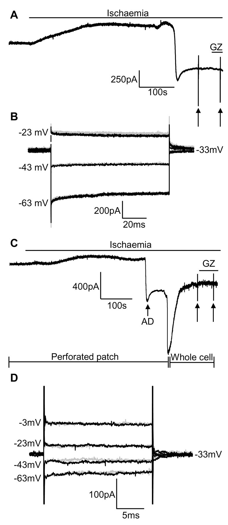
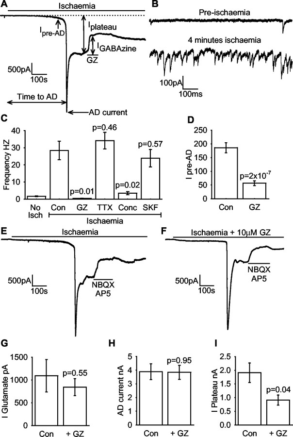
GABA release during cerebral energy deprivation (produced by anoxia or ischemia) has been suggested either to be neuroprotective, because GABA will hyperpolarize neurons and reduce release of excitotoxic glutamate, or to be neurotoxic, because activation of GABA(A) receptors facilitates Cl- entry into neurons and consequent cell swelling. We have used the GABA(A) receptors of hippocampal area CA1 pyramidal cells to sense the rise of [GABA](o) occurring in simulated ischemia. Ischemia evoked, after several minutes, a large depolarization to approximately -20 mV. Before this "anoxic depolarization," there was an increase in GABA release by exocytosis (spontaneous IPSCs). After the anoxic depolarization, there was a much larger, sustained release of GABA that was not affected by blocking action potentials, vesicular release, or the glial GABA transporter GAT-3 but was inhibited by blocking the neuronal GABA transporter GAT-1. Blocking GABA(A) receptors resulted in a more positive anoxic depolarization but decreased cell swelling at the time of the anoxic depolarization. The influence of GABA(A) receptors diminished in prolonged ischemia because glutamate release evoked by the anoxic depolarization inhibited GABA(A) receptor function by causing calcium entry through NMDA receptors. These data show that ischemia releases GABA initially by exocytosis and then by reversal of GAT-1 transporters and that the resulting Cl- influx through GABA(A) receptor channels causes potentially neurotoxic cell swelling.


Figures

Figure 9.
Figure 9.
GABAA receptors are inactivated after the AD in CA1 neurons when the internal calcium buffering of the cell is not altered by whole-cell clamping. A, Recording of a CA1 neuron at –33 mV in the perforated-patch mode, showing no response to the application of GABAzine 2 min after the AD; arrows mark two sets of voltage steps applied before and in GABAzine. B, Current responses to voltage steps applied before GABAzine (black lines) and in GABAzine (gray lines). The two sets of traces superimpose, showing that the lack of response to GABAzine seen at –33 mV in A is not attributable to the chloride reversal potential being close to –33 mV (4 cells had GABAzine applied, and two of these were stepped to different potentials as here). C, D, GABAA receptors remain desensitized after the AD, after going to the whole-cell mode from the perforated-patch mode with a BAPTA-buffered internal solution. C, Cell recorded in the perforated-patch mode at –33 mV; the first large inward deflection is the AD current, and the second is the cell shifting from the perforated to the whole-cell mode (as assessed by the entry of Lucifer yellow into the cell). Application of GABAzine did not affect the plateau current, despite the presence of BAPTA inside the cell. D, Current response to voltage steps applied before GABAzine (black lines) and in GABAzine (gray lines). The two sets of traces superimpose, showing that the lack of response to GABAzine in C is not attributable to the chloride reversal potential shifting to be at –33 mV (done in 4 cells).
Figure 7.
Figure 7.
Activation of GABAA receptors at the time of the AD potentiates cell swelling. A, Schematic diagram of light transmittance measurement. Intracellular organelles scatter light and reduce the transmitted light. When cells swell, the organelles are diluted, reducing scattering and increasing transmittance. B, Light levels transmitted through a slice remained constant until the time of the AD, when there was a sudden increase in transmittance attributed to cellular swelling. The bottom trace shows the extracellularly recorded field potential for correlation of the timing of swelling and of the AD. C, With GABAA receptors blocked, the initial increase in light transmittance (marked by a horizontal arrow) at the time of the AD was reduced, but transmittance continued to increase slowly over a number of minutes. D, The initial light increase at the time of the AD was reduced in GABAzine (8 slices control ischemia solution, 8 interleaved slices with GABAzine). E, The final increase in light transmittance, measured 5 min after the AD, was unchanged in the presence of GABAzine. F, The initial light increase at the time of the AD was reduced by pretreating slices with 0.5 μm concanamycin and applying 100 μm SKF-89976A throughout (14 slices control ischemia, 15 interleaved slices with concanamycin plus SKF-89976A). G, The final increase in light transmittance, measured 5 min after the AD, was unchanged by treatment with concanamycin and SKF-89976A.
Figure 1.
Figure 1.
Ischemia evokes GABA release before and after the AD. A, Current response of a CA1 pyramidal cell to ischemia in the whole-cell voltage clamp mode, with measured parameters marked (dashed line indicates the extrapolated baseline). The plateau current was measured 2 min after the AD; GABAzine(GZ) was then applied to assess GABA release, and the current suppressed was measured 3 min after the AD.B, Expanded regions taken from 1 min before and 4 min after ischemia solution was applied (2 min before the AD), showing that there is an increase in the frequency of spontaneous postsynaptic currents in the buildup to the AD. C, Frequency of spontaneous postsynaptic currents in non-ischemic solution (No Isch; 27 cells) and 4 min after application of normal ischemic solution, or ischemic solution containing 10 μm GABAzine (GZ), 1 μm TTX, or 100 μm SKF-89976A (SKF), or normal ischemic solution in slices pretreated with concanamycin (Conc) (4 cells for each ischemic condition). D, Magnitude of the pre-AD current (1 min before the AD, measured from the pre-ischemic baseline) in control ischemia solution and in ischemia solution containing 10 μm GABAzine.E, Application of 25 μm NBQX and 50 μm AP5 shows that about half of the plateau current after the AD is generated by glutamate receptors.F, For ischemia in the presence of GABAzine throughout, NBQX plus AP5 blocks almost all of the plateau current. G, The size of the glutamate receptor-mediated current (measured as in A for the GABAzine-blocked current) was not affected by blocking GABAA receptors (6 interleaved cells in each group).H, The amplitude of the AD current was not affected by blocking GABAA receptors. I, The plateau current was halved in the presence of GABAzine. All data are at –33 mV.
Figure 6.
Figure 6.
Blocking GABAA receptors alters the potential reached during the response of CA1 pyramidal cells to ischemia. A, Current-clamp recording in perforated patch configuration. On application of ischemia solution, the cell deploarizes by a few millivolts (thick arrow) and then hyperpolarizes (thin arrow), then the AD occurs after a few minutes. Large vertical thin lines are action potentials. B, As in A, but with GABAA receptors blocked by GABAzine. C, The resting potentials of the cells studied without (control) and with GABAzine were not significantly different before ischemia. D, Cells in GABAzine deploarized more at the time of the AD (6 cells control, 5 cells GABAzine).
Figure 8.
Figure 8.
GABAA receptors become nonfunctional after the AD as a result of Ca2+ entry through NMDA receptors. A, Response to ischemia of a cell whole-cell clamped with BAPTA as the internal calcium buffer: GABAzine reduces the inward plateau current after the AD. B, Response to ischemia of a cell whole-cell clamped with EGTA as the calcium buffer: GABAzine has no effect on the plateau current. C, Mean GABAzine-blockable current after the AD when cells were dialyzed with BAPTA (6 cells) or with EGTA internal solution (8 interleaved cells). D, Using BAPTA, a cell responds to exogenous 1 mm GABA after the AD. E, Using EGTA a cell does not respond to GABA after the AD. F, Mean GABA-evoked current after the AD when cells were dialyzed with BAPTA (5 cells) or with EGTA internal solution (5 cells). G, The amplitude of the plateau current after the AD was halved when EGTA was used as the calcium buffer (10 cells dialyzed with EGTA, 10 interleaved cells with BAPTA). H, Blocking NMDA receptors with 50 μm MK-801 throughout ischemia in a cell whole-cell clamped with EGTA internal solution allows a GABAzine-blockable current to remain after the AD. I, MK-801 delayed the AD (7 EGTA cells in control solution, 8 interleaved EGTA cells with MK-801). J, MK-801 decreased the amplitude of the AD current. K, Mean GABAzine-blocked current after the AD with EGTA internal was rescued by MK-801. All data are at –33 mV.
Figure 2.
Figure 2.
GABA release after the AD is not action potential evoked and is not by exocytosis. A, In the presence of 1 μm TTX to block action potentials during application of ischemia solution, the current response of CA1 pyramidal neurons to ischemia was unchanged (compare Fig. 1 A). B, The size of the post-AD GABAzine-blocked current was not significantly different in the presence and absence of TTX (9 control cells, 6 interleaved TTX cells). C, Depleting vesicles of GABA using concanamycin abolished IPSCs evoked in CA1 pyramidal neurons by electrical stimulation (in the presence of 25 μm NBQX and 50 μm AP5 to block glutamate receptors), showing that vesicle depletion had been successful [7 control (Con) cells, 7 concanamycin (Concan) cells; p < 0.05 for all stimulation voltages above 10 V]. D, Blocking vesicular GABA release had little effect on the response of CA1 pyramidal neurons to ischemia (compare Fig. 1 A). E, Blocking vesicular GABA release slightly prolonged the time to the AD. F, Blocking vesicular GABA release had no significant effect on the size of the AD current. G, The amplitude of the GABAzine-blockable current after the AD was not affected by blocking vesicular release (10 cells in control ischemia solution, 8 interleaved cells pretreated with concanamycin for E–G). All data are at –33 mV.
Figure 3.
Figure 3.
GABA release after the AD is mostly by reversed operation of the GAT-1 transporter. A, SKF-89976A (100 μm) only slightly reduced the response to the nontransported GABA analog THIP (20 μm, in non-ischemic solution). Mean data from five cells for the size of the THIP response in SKF-89976A normalized to its response in the absence of 100 μm SKF-89976A (602 ± 24 pA). B, The ratio of the responses of a pyramidal cell to GABA (10 μm) and to THIP (in non-ischemic solution) was increased by 100 μm SKF-89976A. C, Mean ratio of currents as in B (8 cells). D–G, SKF-89976A reduces ischemia-evoked GABA release after the AD. D, In the presence of SKF-89976A throughout ischemia to block GAT-1, the GABAzine-evoked current after the AD was reduced, showing a reduction of GABA release. E, The time of the AD was not altered when GAT-1 was blocked with SKF-89976A. F, The amplitude of the AD current was not affected by SKF-89976A. G, The GABAzine-blockable current was reduced by 70% when GAT-1 was blocked (7 cells studied in control ischemia solution, 8 interleaved cells with SKF-89976A). All data are at –33 mV.
Figure 4.
Figure 4.
β-Alanine preloading blocks the glial GABA transporter GAT-3 but does not reduce ischemia-evoked GABA release. A, Response of a pyramidal cell in an untreated slice in non-ischemic solution to GABA (10 μm) and to the nontransported GABA analog THIP (20 μm). B, As in A, but in a β-alanine-loaded slice. C, The size of the THIP response in A and B was not affected by β-alanine loading. D, The ratio of the GABA response to the THIP response increased 2.5-fold in β-alanine-loaded slices (data from 6 control and 6 β-alanine-treated interleaved slices for C and D). E, After β-alanine loading, the response to ischemia was little affected (compare Fig. 1 A). F, The time to the AD was slightly prolonged in β-alanine-loaded slices (data from 7 control and 6 β-alanine-loaded interleaved slices for F–H). G, The amplitude of the AD current was not affected by β-alanine. H, The size of the GABAzine-blockable current after the AD was not affected by β-alanine preloading. All data are at –33 mV.
Figure 5.
Figure 5.
Blocking all ionotropic glutamate receptors does not prevent GABA release during ischemia. A, Response of a cell to ischemia in the presence of 25 μm NBQX, 50 μm d-AP5, 50 μm MK-801, and 100 μm 7-chlorokynurenic acid to block glutamate receptors. The ischemic response was delayed, but a slow inward current developed after 10 min that was completely blocked by GABAzine, showing it to be mediated by GABAA receptor activation. Increased noise on the trace is spontaneous synaptic currents. B, With GAT-1 blocked throughout with 100 μm SKF-89976A, a GABAA-mediated inward current still developed but the amplitude was greatly reduced, whereas spontaneous events were still seen before the AD. C, The amplitude of the GABAzine-blockable current was reduced by 68% when GAT-1 was blocked (8 cells studied in control solution, 5 interleaved cells with 100 μm SKF-89976A). All data are at –33 mV.

References

    1. Aika Y, Ren JQ, Kosaka K, Kosaka T (1994) Quantitative analysis of GABA-like-immunoreactive and parvalbumin-containing neurons in the CA1 region of the rat hippocampus using a stereological method, the disector. Exp Brain Res 99: 267–276. - PubMed
    1. Akaike N (1996) Gramicidin perforated patch recording and intracellular chloride activity in excitable cells. Prog Biophys Mol Biol 65: 251–264. - PubMed
    1. Alicke B, Schwartz-Bloom RB (1995) Rapid down-regulation of GABAA receptors in the gerbil hippocampus following transient cerebral ischaemia. J Neurochem 65: 2808–2811. - PubMed
    1. Ankarcrona M, Dypbukt JM, Bonfoco E, Zhivotovsky B, Orrenius S, Lipton SA, Nicotera P (1995) Glutamate-induced neuronal death: a succession of necrosis or apoptosis depending on mitochondrial function. Neuron 15: 961–973. - PubMed
    1. Attwell D, Barbour B, Szatkowski M (1993) Nonvesicular release of neurotransmitter. Neuron 11: 401–407. - PubMed

Publication types

MeSH terms

LinkOut - more resources