. 2004 Apr 14;32(6):e58.
doi: 10.1093/nar/gnh063.

Peptide-linked molecular beacons for efficient delivery and rapid mRNA detection in living cells

Affiliations

Peptide-linked molecular beacons for efficient delivery and rapid mRNA detection in living cells

Nitin Nitin et al. Nucleic Acids Res. .

Abstract

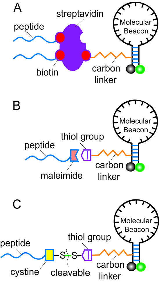
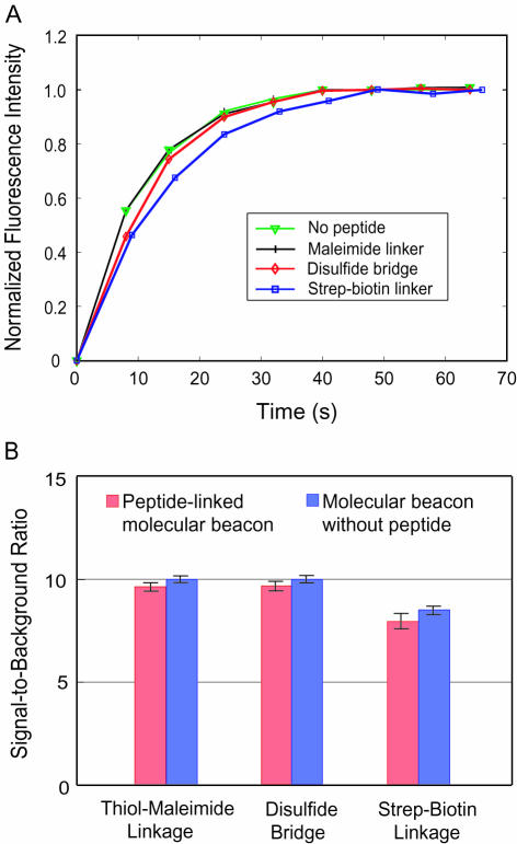
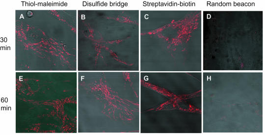
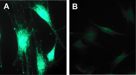
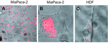
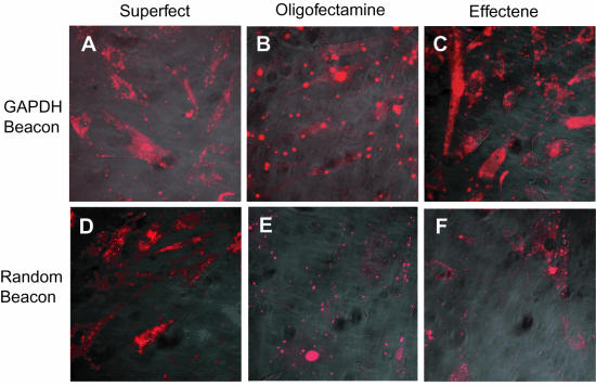
Real-time visualization of specific endogenous mRNA expression in vivo has the potential to revolutionize medical diagnosis, drug discovery, developmental and molecular biology. However, conventional liposome- or dendrimer-based cellular delivery of molecular probes is inefficient, slow, and often detrimental to the probes. Here we demonstrate the rapid and sensitive detection of RNA in living cells using peptide-linked molecular beacons that possess self-delivery, targeting and reporting functions. We conjugated the TAT peptide to molecular beacons using three different linkages and demonstrated that, at relatively low concentrations, these molecular beacon constructs were internalized into living cells within 30 min with nearly 100% efficiency. Further, peptide-based delivery did not interfere with either specific targeting by or hybridization-induced fluorescence of the probes. We could therefore detect human GAPDH and survivin mRNAs in living cells fluorescently, revealing intriguing intracellular localization patterns of mRNA. We clearly demonstrated that cellular delivery of molecular beacons using the peptide-based approach has far better performance compared with conventional transfection methods. The peptide-linked molecular beacons approach promises to open new and exciting opportunities in sensitive gene detection and quantification in vivo.


Figures

Figure 1
Figure 1
A schematic illustration of three different conjugation schemes for linking the delivery peptide to molecular beacons. (A) The streptavidin–biotin linkage in which a molecular beacon is modified by introducing a biotin-dT to the quencher arm of the stem through a carbon-12 spacer. The biotin-modified peptides are linked to the modified molecular beacon through a streptavidin molecule, which has four biotin-binding sites. (B) The thiol–maleimide linkage in which the quencher arm of the molecular beacon stem is modified by adding a thiol group which can react with a maleimide group placed to the C terminus of the peptide to form a direct, stable linkage. (C) The cleavable disulfide bridge in which the peptide is modified by adding a cysteine residue at the C terminus which forms a disulfide bridge with the thiol-modified molecular beacon. This disulfide bridge design allows the peptide to be cleaved from the molecular beacon by the reducing environment of the cytoplasm.
Figure 2
Figure 2
Probe–target hybridization kinetics of unmodified and peptide-linked molecular beacons. (A) Normalized fluorescence intensity as a function of time for unmodified molecular beacons, and for the three types of peptide-linked molecular beacons. Note that the probe–target hybridization kinetic curves of peptide-linked molecular beacons with the thiol–maleimide linkage (black curve) and unmodified molecular beacons (green curve) are almost identical. (B) S/B ratios (red) of probe–target hybridization for peptide-linked molecular beacons with different conjugation methods. The S/B ratios for thiol-modified and biotin-modified molecular beacons (blue) without peptide are also shown for comparison. The hybridization kinetics and S/B ratio did not change much, indicating that the functionality of the molecular beacons was not impaired by peptide conjugation.
Figure 3
Figure 3
Detection of GAPDH mRNA in HDF cells using 0.5 µM of peptide-linked molecular beacons. (AC) Fluorescence images of HDF cells after 30 min of incubation with TAT-peptide conjugated, GAPDH-targeting molecular beacons, with (A) thiol–maleimide, (B) disulfide bridge and (C) streptavidin–biotin linkages. (D) With peptide-linked random-sequence molecular beacons, there was essentially no detectable signal 30 min after delivery. (EG) Fluorescence images of HDF cells after 60 min of incubation of peptide-linked, GAPDH-targeting molecular beacons with (E) thiol–maleimide, (F) disulfide bridge and (G) streptavidin–biotin linkages. The signal level of peptide-linked random-sequence molecular beacons after 60 min is shown in (H). Clearly, the fluorescence signal level did not increase when incubation time was doubled, indicating that most of the molecular beacons internalized within the first 30 min. Note the intriguing GAPDH mRNA localization patterns shown in (A–C) and (E–G).
Figure 4
Figure 4
Control studies using fluorescence in situ hybridization (FISH). (A) Detection of GAPDH mRNA in fixed HDF cells. The filamentous localization pattern of GAPDH mRNA was similar to that revealed in living cell assays, although the signal was not as sharp. Note the diffused fluorescence signal in cell nuclei. (B) A negative control study of the FISH assay using fluorescently labeled linear Poly-A probes resulted in very low background.
Figure 5
Figure 5
Detection of survivin mRNA in live HDF and MiaPaca-2 cells. (A) A strong fluorescence signal was observed in MiaPaca-2 cells after 60 min of incubation with peptide-linked molecular beacons. Note that essentially all cells showed fluorescence. (B) Survivin mRNA molecules in MiaPaca-2 cells seemed to be concentrated near one side of the cell nucleus of HDF cells. (C) Only a very low fluorescence signal can be observed in HDF cells, but with a survivin mRNA localization pattern.
Figure 6
Figure 6
Cellular delivery using conventional transfection methods. (AC) Fluorescence signal in HDF cells after 3.5 h transfection of unmodified GAPDH-targeting molecular beacons with (A) Superfect, (B) Oligofectamine and (C) Effectene. Note the concentrated ‘bright spots’ in both cytoplasm and nucleus. (DF) Similar fluorescence signal levels were observed after 3.5 h delivery of random-sequence molecular beacons with (D) Superfect, (E) Oligofectamine and (F) Effectene. The resulting ‘bright spots’ in HDF cells indicate that the fluorescence signals in (A–F) were largely due to molecular beacon degradation.

References

    1. Karge W.H. 3rd, Schaefer,E.J. and Ordovas,J.M. (1998) Quantification of mRNA by polymerase chain reaction (PCR) using an internal standard and a nonradioactive detection method. Methods Mol. Biol., 110, 43–61. - PubMed
    1. Jung R., Soondrum,K. and Neumaier,M. (2000) Quantitative PCR. Clin. Chem. Lab. Med., 38, 833–836. - PubMed
    1. Brown P.O. and Botstein,D. (1999) Exploring the new world of the genome with DNA microarrays. Nature Genet., 21, 33–37. - PubMed
    1. DeRisi J.L. and Iyer,V.R. (1999) Genomics and array technology. Curr. Opin. Oncol., 11, 76–79. - PubMed
    1. Femino A.M., Fay,F.S., Fogarty,K. and Singer,R.H. (1998) Visualization of single RNA transcripts in situ. Science, 280, 585–590. - PubMed

Publication types

MeSH terms