. 2004 Apr;134(4):1427-38.
doi: 10.1104/pp.103.036780.

Phosphorylation of the amino terminus of maize sucrose synthase in relation to membrane association and enzyme activity

Affiliations

Phosphorylation of the amino terminus of maize sucrose synthase in relation to membrane association and enzyme activity

Shane C Hardin et al. Plant Physiol. 2004 Apr.

Abstract

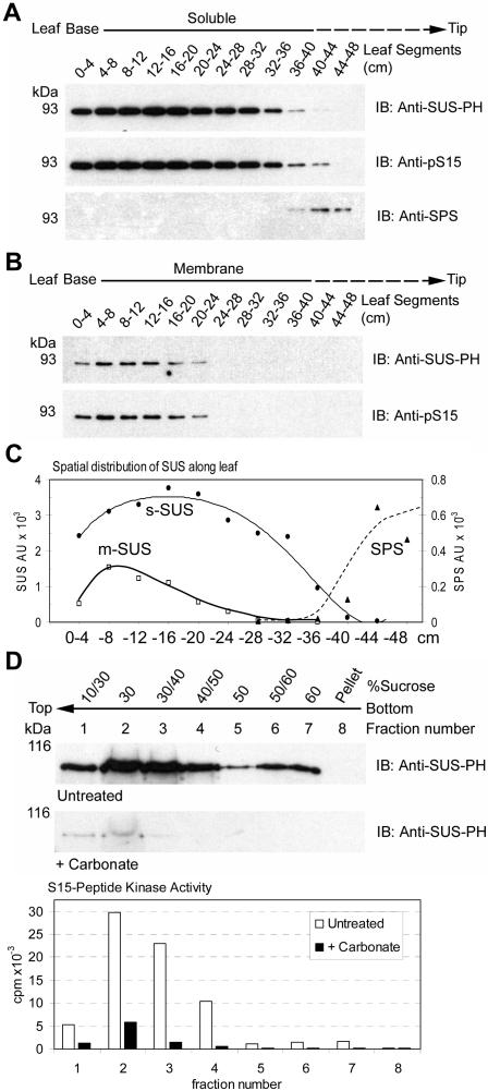
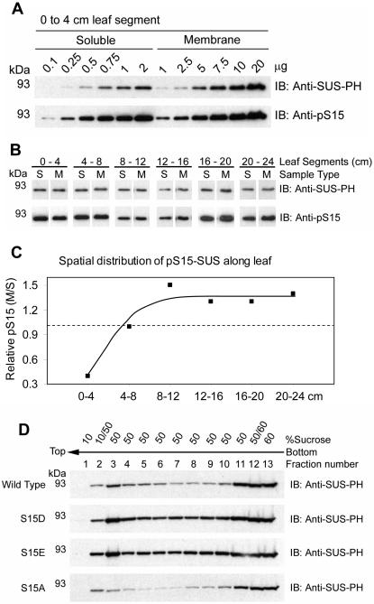
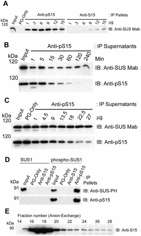
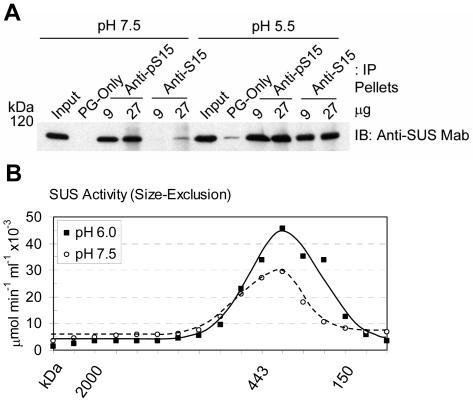
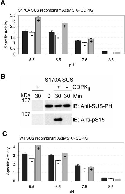
Sucrose synthase (SUS) is phosphorylated on a major, amino-terminal site located at Ser-15 (S15) in the maize (Zea mays) SUS1 protein. Site- and phospho-specific antibodies against a phosphorylated S15 (pS15) peptide allowed direct analysis of S15 phosphorylation in relation to membrane association. Immunoblots of the maize leaf elongation zone, divided into 4-cm segments, demonstrated that the abundance of soluble (s-SUS) and membrane (m-SUS) SUS protein showed distinct positional profiles. The content of m-SUS was maximal in the 4- to 8-cm segment where it represented 9% of total SUS and occurred as a peripheral membrane protein. In contrast, s-SUS was highest in the 12- to 16-cm segment. Relative to s-SUS, m-SUS was hypophosphorylated at S15 in the basal 4 cm but hyperphosphorylated in apical segments. Differing capabilities of the anti-pS15 and anti-S15 peptide antibodies to immunoprecipitate SUS suggested that phosphorylation of S15, or exposure of unphosphorylated SUS to slightly acidic pH, altered the structure of the amino terminus. These structural changes were generally coincident with the increased sucrose cleavage activity that occurs at pH values below 7.5. In vitro S15 phosphorylation of the S170A SUS protein by a maize calcium-dependent protein kinase (CDPK) significantly increased sucrose cleavage activity at low pH. Collectively, the results suggest that (1) SUS membrane binding is controlled in vivo; (2) relative pS15 content of m-SUS depends on the developmental state of the organ; and (3) phosphorylation of S15 affects amino-terminal conformation in a way that may stimulate the catalytic activity of SUS and influence membrane association.


Figures

Figure 1.
Figure 1.
The distribution of SUS between the soluble and membrane phases is developmentally regulated in maize leaves. A, Equal amounts of total soluble protein extracts (1 μg) isolated from sequential 4-cm segments of elongating maize leaves were probed on immunoblots (IB) with the antibodies listed on the right of each panel. The positions of PAGE molecular mass markers are shown in kilodaltons on the left of each panel. B, Equal amounts of total membrane phase protein extracts (1 μg) isolated from sequential 4-cm segments of maize leaves were probed on immunoblots as in (A). C, Densitometry of immunoblots in A and B showing the distribution of anti-SUS-PH and anti-SPS antibody reactive proteins along the leaf. Arbitrary units (AU) are shown. D, Membrane preparations from maize leaves were untreated or treated with carbonate and subjected to flotation on discontinuous Suc gradients. Immunoblots (IB) were performed on the subsequent fractions with the antibodies listed to the right of each panel. S15-peptide kinase activities (cpm × 10−3) within the untreated (white bars) and carbonate-treated (black bars) fractions is also shown.
Figure 2.
Figure 2.
Relative phosphorylation of S15 is variable along elongating maize leaves. A, Increasing amounts of total soluble (0.1–2 μg) and membrane phase (1–20 μg) protein extracts isolated from the basal 0- to 4-cm segment of maize leaves were probed on immunoblots (IB) with the antibodies listed on the right of each panel. The positions of PAGE molecular mass markers are shown in kilodaltons on the left of each panel. B, Representative data of single load comparisons extracted from 12 immunoblots performed as in A that included extracts isolated from sequential 4-cm segments of maize leaves covering the region from 0 to 24 cm. C, Relative pS15 content of m-SUS compared to s-SUS in different regions of the elongation zone. Densitometry of immunoblots, such as the dilution series shown in A, was used to determine the slopes of total protein versus antibody signal (AU). These values were used to determine the relative phosphorylation of S15 (per unit SUS protein) in the membrane and soluble phase for each 4-cm segment over the basal 24 cm of the leaf and are depicted as a ratio. Values greater than 1.0 (dashed line) indicate hyperphosphorylation of S15 in m-SUS relative to s-SUS while values less than 1.0 indicate hypophosphorylation of S15. D, Membrane preparations recovered from the SUS recombinants listed to the left of each panel were subjected to flotation on discontinuous Suc gradients. Immunoblots (IB) were performed on the subsequent fractions with the antibodies listed to the right of each panel.
Figure 3.
Figure 3.
Phosphorylation of S15 affects the conformation of the amino terminus of SUS. A, Immunoprecipitation (IP) of native s-SUS was performed with blank Protein-G beads (PG-only) or with the antibodies listed at the top of the figure. Immunoblots (IB) were performed on the IP pellets with the antibody listed to the right of the figure. The positions of PAGE molecular mass markers are shown in kilodaltons on the left of the figure. B, Immunoprecipitation (IP) of native s-SUS was performed for increasing amounts of time with 13.5 μg of the antibody listed at the top of the figure. Immunoblots (IB) were performed on the IP supernatants with the antibodies listed to the right of each panel. The positions of PAGE molecular mass markers are shown in kilodaltons (kD) on the left of each panel. C, Immunoprecipitation (IP) of native s-SUS was performed with blank Protein-G beads (PG-only) or with increasing amounts of the antibody listed at the top of the figure for 120 min. Immunoblots (IB) were performed on the IP supernatants with the antibodies listed to the right of each panel. D, Immunoprecipitation (IP) of recombinant wild-type SUS1 or CDPKII-phosphorylated SUS1 (phospho-SUS1 including pS15 and pS170) was performed with blank Protein-G beads (PG-only) or with the antibodies listed at the top of the figure. Immunoblots (IB) were performed on the IP pellets with the antibodies listed to the right of each panel. E, Native s-SUS protein was resolved by anion-exchange chromatography and immunoblot (IB) analysis performed on the SDS-denatured form with anti-S15 antibodies.
Figure 4.
Figure 4.
Low pH affects the conformation of the amino terminus of SUS without affecting oligomerization state of the native protein. A, Immunoprecipitation (IP) of native s-SUS was performed at pH 7.5 or pH 5.5 with blank Protein-G beads (PG-only) or with the antibodies listed at the top of the figure. Immunoblots (IB) were performed on the IP pellets with the antibody listed to the right of the figure. The positions of PAGE molecular mass markers are shown in kilodaltons on the left of the figure. B, Sucrose cleavage activity (μmol UDP-Glc min−1 mL−1 × 10⁻³) of native s-SUS resolved by size-exclusion chromatography at pH 6.0 (▪, solid line) or pH 7.5 (○, dashed line). The elution positions of molecular mass standards are shown in kilodaltons (kD) under the graph.
Figure 5.
Figure 5.
Phosphorylation affects the Suc cleavage activity of SUS. A, Specific Suc cleavage activities (μmol UDP-Glc min−1 mg−1) at pH 5.5, 6.5, 7.5, and 8.5 of S170A SUS1 recombinants at time zero (black bars) or after a 30-min incubation in vitro in the presence (+, gray bars) or absence (−, white bars) of CDPKII. B, Immunoblot (IB) analyses of CDPKII and S170A SUS1 recombinants at time zero or after a 30-min incubation in vitro in the presence (+) or absence (−) of CDPKII, with the antibodies listed to the right of each panel. The positions of PAGE molecular mass markers are shown in kilodaltons on the left of the figure. C, Specific Suc cleavage activities (μmol UDP-Glc min−1 mg−1) at pH 5.5, 6.5, 7.5, and 8.5 of wild-type SUS1 recombinants at time zero (black bars) or after a 30-min incubation in vitro in the presence (+, gray bars) or absence (−, white bars) of CDPKII.

References

    1. Ahola T, Lampio A, Auvinen P, Kaariainen L (1999) Semliki Forest virus mRNA capping enzyme requires association with anionic membrane phospholipids for activity. EMBO J 18: 3164–3172 - PMC - PubMed
    1. Amor Y, Haigler CH, Johnson S, Wainscott M, Delmer DP (1995) A membrane-associated form of sucrose synthase and its potential role in synthesis of cellulose and callose in plants. Proc Natl Acad Sci USA 92: 9353–9357 - PMC - PubMed
    1. Anguenot R, Yelle S, Nguyen-Quoc B (1999) Purification of tomato sucrose synthase phosphorylated isoforms by Fe(III)-immobilized metal affinity chromatography. Arch Biochem Biophys 365: 163–169 - PubMed
    1. Azama K, Abe S, Sugimoto H, Davies E (2003) Lysine-containing proteins in maize endosperm: a major contribution from cytoskeleton-associated carbohydrate-metabolizing enzymes. Planta 217: 628–638 - PubMed
    1. Bell-Parikh LC, Eipper BA, Mains RE (2001) Response of an integral granule membrane protein to changes in pH. J Biol Chem 276: 29854–29863 - PubMed

Publication types

MeSH terms

LinkOut - more resources