. 2004 Apr 27;101(17):6490-5.
doi: 10.1073/pnas.0308684101. Epub 2004 Apr 14.

Lactosylceramide recruits PKCalpha/epsilon and phospholipase A2 to stimulate PECAM-1 expression in human monocytes and adhesion to endothelial cells

Affiliations

Lactosylceramide recruits PKCalpha/epsilon and phospholipase A2 to stimulate PECAM-1 expression in human monocytes and adhesion to endothelial cells

NanLing Gong et al. Proc Natl Acad Sci U S A. .

Abstract

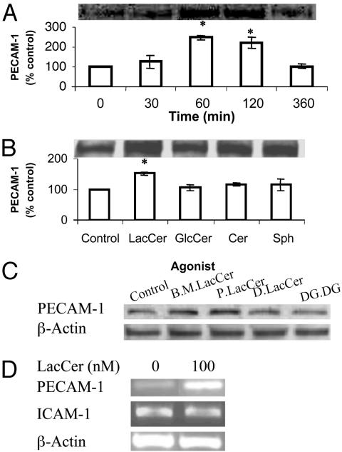
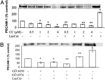
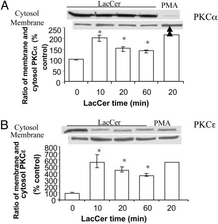
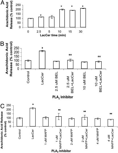
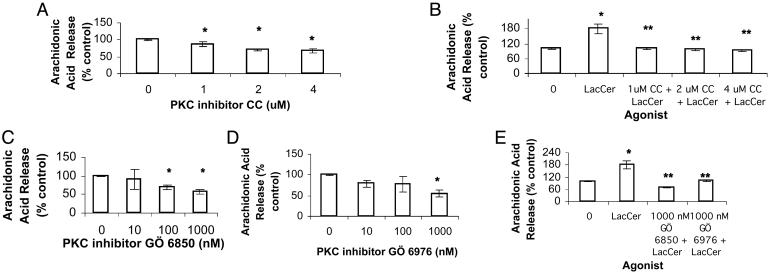
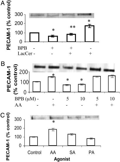
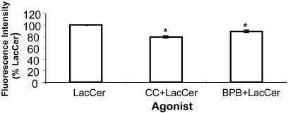
Despite the importance of platelet/endothelial cell adhesion molecule-1 (PECAM-1, CD31) in the adhesion and diapedesis of monocytes/lymphocytes, little is known about the mechanisms by which it is regulated. We explored the role of a glycosphingolipid, lactosylceramide (LacCer), in modulating PECAM-1 expression and cell adhesion in human monocytes. We observed that LacCer specifically exerted a time-dependent increase in PECAM-1 expression in U-937 cells. Maximal increase in PECAM-1 protein occurred after incubation with LacCer for 60 min. LacCer activated PKCalpha and -epsilon by translocating them from cytosol to membrane. This was accompanied by the activation of phospholipase A(2) (PLA(2)) and the increase of cell adhesion, which were abrogated by chelerythrine chloride, 2-[1-(3-dimethylaminopropyl)-1H-indol-3-yl]-3-(1H-indol-3-yl)-maleimide and 12-(2-cyanoethyl)-6,7,12,13-tetrahydro-13-methyl-5-oxo-5H-indolo(2,3-a)pyrrolo(3,4-c)-carbazole (GO 6976) (PKC inhibitors). Similarly, bromoenol lactone (a Ca(2+)-independent PLA(2) inhibitor) and methyl arachidonyl fluorophosphonate (an inhibitor of cytosolic PLA(2) and Ca(2+)-independent PLA(2)) inhibited LacCer-induced PLA(2) activity. Bromophenacyl bromide (a PLA(2) inhibitor) abrogated LacCer-induced PECAM-1 expression, and this was bypassed by arachidonic acid. Furthermore, the arachidonate-induced up-regulation of PECAM-1 was abrogated by indomethacin [a cyclooxygenase (COX)-1 and -2 inhibitor] or N-[2-(cyclohexyloxy)-4-nitrophenyl]-methanesulfonamide (a COX-2 inhibitor) but not nordihydroguaiaretic acid (a lipoxygenase inhibitor). In sum, PKCalpha/epsilon are the primary targets for the activation of LacCer. Downstream activation of intracellular Ca(2+)-independent PLA(2) and/or cytosolic PLA(2) results in the production of arachidonic acid, which in turn serves as a precursor for prostaglandins that subsequently stimulate PECAM-1 expression and cell adhesion. These findings may be relevant in explaining the role of LacCer in the regulation of PECAM-1 and related pathophysiology.


Figures

Fig. 1.
Fig. 1.
Effect of 100 nM bovine brain LacCer (Sigma) (A), its metabolites (B), and bovine milk (B.M.) LacCer, palmitoyl LacCer (Matreya; P.LacCer), dihydro LacCer (D.LacCer), and digalactosyl diglyceride (DG.DG.) (C) on PECAM-1 protein expression in U-937 cells. Western immunoblot analysis and densitometric scanning of PECAM-1 level in cells in serum-free RPMI medium 1640, treated with indicated agonists for indicated periods of time in A, and for 1 h in B and C. (D) Effect of LacCer on PECAM-1 and ICAM-1 mRNA transcription in U-937 cells. Total RNA was extracted from cells (1 × 10⁷) incubated with 100 nM LacCer for 1 h and an equal amount of total RNA was subjected to semiquantitative RT-PCR. β-Actin served as an internal control to verify the equal amount of total cDNA used for each sample (P < 0.05 vs. control).
Fig. 2.
Fig. 2.
Effect of PKC inhibitors chelerythrine chloride (CC; A) and GÖ 6976 and GÖ 6850 (B) on LacCer-mediated PECAM-1 protein expression in U-937 cells. Western immunoblot analysis and densitometric scanning of PECAM-1 in cells treated with various concentrations of chelerythrine chloride (CC) for 2 h (A) and 10 nM GÖ 6976 or GÖ 6850 for3h(B) followed with and without 100 nM LacCer for 1 h. *, P < 0.05 vs. control; **, P < 0.05 vs. LacCer; n = 3.
Fig. 3.
Fig. 3.
Membrane and cytosol distribution of PKC isozymes α (A) and ε (B)in U-937 cells. Western immunoblot analysis and densitometric scanning of PKCα and -ε in membrane and cytosol of cells treated with and without 100 nM LacCer for the indicated time in serum-free RPMI medium 1640. Phorbol 12-myristate 13-acetate treatment (0.1 μM, 20 min) of U-937 cells served as a positive control. *, P < 0.05 vs. control; n = 3.
Fig. 4.
Fig. 4.
(A) LacCer activates PLA2 in U-937 cells in a time-dependent manner. Cells (1 × 10⁶) were labeled with [3H]arachidonic acid for 30 min, washed with serum-free HBSS serially four times, and then incubated with 100 nM LacCer for various time points (0, 2.5, 5, 10, 15, and 30 min). Next, radioactivity in the supernatant was measured. (B and C) PLA2 inhibitors abrogate increased arachidonic acid release from LacCer-treated [3H]arachidonic acid-labeled U-937 cells. [3H]arachidonic acid-labeled U-937 cells were treated with various concentrations of iPLA2 inhibitor bromoenol lactone (BEL; 2.5 and 10 μM) (B), iPLA2 and cPLA2 inhibitor methyl arachidonyl fluorophosphonate (MAFP; 1, 2, and 4 μM) (C) for 30 min at 37°C, followed with and without 100 nM LacCer for 30 min. Data represent mean ± SD of three different experiments in duplicates. *, P < 0.05 vs. control; **, P < 0.05 vs. LacCer.
Fig. 5.
Fig. 5.
PKC inhibitors abrogate LacCer-mediated PLA2 activation. Arachidonic acid release assay was done. [3H]Arachidonic acid-labeled U-937 cells were treated with chelerythrine chloride (CC) (A and B), GÖ 6850 and GÖ 6976 (CE) for 30 min followed with and without 100 nM LacCer for 30 min at 37°C. Data represent mean ± SD of three different experiments in duplicates. *, P < 0.05 vs. control; **, P < 0.05 vs. LacCer.
Fig. 6.
Fig. 6.
Effect of PLA2 inhibitor BPB (A), arachidonic acid (AA; B), and other fatty acids (C) on PECAM-1 protein expression in U-937 cells. Western immunoblot analysis and densitometric scanning of PECAM-1 in cells treated with 100 nM LacCer for 1 h with and without pretreatment with 10 μM BPB for 30 min (A), 10 μM AA for 1 h with and without pretreatment with BPB for 30 min (B), 10 μM AA, stearic acid (SA), and palmitic acid (PA) for 1 h (C). *, P < 0.05 vs. control; **, P < 0.05 vs. LacCer; n = 3.
Fig. 7.
Fig. 7.
LacCer-mediated increase in cell adhesion is abrogated by PKC inhibitor and PLA2 inhibitor. U-937 cells were treated with 100 nM LacCer with and without PKC inhibitor chelerythrine chloride (CC; 4 μM) and PLA2 inhibitor BPB (5 μM) for 1 h. Adhesion of each category of U-937 cells to HUVECs was done as described in Supporting Methods. Triplicates were used for each treatment. The average fluorescence intensity for the group treated with 100 nM LacCer was 664.5 ± 4.2. It was significantly higher than that of the control (595.5 ± 1.4). Data were plotted by using the fluorescence intensity of LacCer as 100% and represent mean ± SD of two independent experiments. *, P < 0.05 vs. LacCer.

References

    1. Minehata, K., Mukouyama, Y. S., Sekiguchi, T., Hara, T. & Miyajima, A. (2002) Blood 99, 2360-2368. - PubMed
    1. Shih, S. C., Robinson, G. S., Perruzzi, C. A., Calvo, A., Desai, K., Green, J. E., Ali, I. U., Smith, L. E. & Senger, D. R. (2002) Am. J. Pathol. 161, 35-41. - PMC - PubMed
    1. Serebruany, V. L., Murugesan, S. R., Pothula, A., Atar, D., Lowry, D. R., O'Connor, C. M. & Gurbel, P. A. (1999) Eur. J. Heart Fail. 1, 243-249. - PubMed
    1. Serebruany, V. L., Murugesan, S. R., Pothula, A., Semaan, H. & Gurbel, P. A. (1999) Cardiology 91, 50-55. - PubMed
    1. Blann, A. D., Devine, C., Amiral, J. & McCollum, C. N. (1998) Blood Coagul. Fibrinolysis 9, 479-484. - PubMed

Publication types

MeSH terms