. 2004 Apr 27;101(17):6542-7.
doi: 10.1073/pnas.0308637101. Epub 2004 Apr 14.

Human centromere repositioning "in progress"

Affiliations

Human centromere repositioning "in progress"

David J Amor et al. Proc Natl Acad Sci U S A. .

Abstract

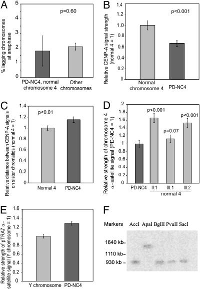
Centromere repositioning provides a potentially powerful evolutionary force for reproductive isolation and speciation, but the underlying mechanisms remain ill-defined. An attractive model is through the simultaneous inactivation of a normal centromere and the formation of a new centromere at a hitherto noncentromeric chromosomal location with minimal detrimental effect. We report a two-generation family in which the centromeric activity of one chromosome 4 has been relocated to a euchromatic site at 4q21.3 through the epigenetic formation of a neocentromere in otherwise cytogenetically normal and mitotically stable karyotypes. Strong epigenetic inactivation of the original centromere is suggested by retention of 1.3 megabases of centromeric alpha-satellite DNA, absence of detectable molecular alteration in chromosome 4-centromereproximal p- and q-arm sequences, and failure of the inactive centromere to be reactivated through extensive culturing or treatment with histone deacetylase inhibitor trichostatin A. The neocentromere binds functionally essential centromere proteins (CENP-A, CENP-C, CENP-E, CENP-I, BUB1, and HP1), although a moderate reduction in CENP-A binding and sister-chromatid cohesion compared with the typical centromeres suggests possible underlying structural/functional differences. The stable mitotic and meiotic transmissibility of this pseudodicentric-neocentric chromosome in healthy individuals and the ability of the neocentric activity to form in a euchromatic site in preference to a preexisting alphoid domain provide direct evidence for an inherent mechanism of human centromere repositioning and karyotype evolution "in progress." We discuss the wider implication of such a mechanism for meiotic drive and the evolution of primate and other species.


Figures

Fig. 1.
Fig. 1.
(A) Pedigree of family carrying PD-NC4, first detected in III:2. Haplotype bars derived from the microsatellite linkage screen indicate that the allele containing the neocentromere (black bar) was derived from the paternal grandfather (I:1) of the proband. (B) The PD-NC4 (Left) and normal chromosome 4 (Right) from G-banded karyotype of individual III:2. The primary constriction has relocated to 4q21.3 without any change in banding pattern. (C) BAC probes used to characterize the PD-NC4 neocentromere (NC) (Left) and alphoid centromere (Right). The neocentromere domain was defined by combined immunofluorescence and FISH to a region of 1.5 Mb bordered by BACs RP11–209g6 and RP11–204i22 (see also Fig. 2D). All 12 BACs flanking the alphoid centromere hybridized with equal intensity to the PD-NC4 and the normal chromosome 4, excluding a deletion of these regions.
Fig. 2.
Fig. 2.
(A) FISH with chromosome 4-specific α-satellite probe (p4n1/4) showing hybridization to the centromere of the normal chromosome 4 (Left) and the inactive centromere (solid arrow) but not the neocentromere (open arrow) of PD-NC4 (Right). (B) FISH with pan-α-satellite probe (pTRA7) showing hybridization to the inactive centromere (solid arrow) of PD-NC4 and the centromere of all other chromosomes but not the PD-NC4 neocentromere (open arrow). (Inset) Combined immunofluorescence and FISH (immuno-FISH) on PD-NC4 using anti-CENP-A antibody (red) and FISH wish pTRA7 (green). (C) Immunofluorescence using anti-CENP-A (green) and anti-CENP-B (red). The CENP-A signal is reduced in strength compared with other centromeres (see also Fig. 3B). (Inset) Immunofluorescence on PD-NC4 using anti-HP1α antibody (red) followed by FISH using a BAC probe from the neocentromere site to identify PD-NC4 (picture not shown), showing the presence of HP1α at both the inactive alphoid centromere and the neocentromere. (D) Localization of the neocentromere site using BAC probes (see Fig. 1C; green) and anticentromere antibody (CREST6, red), showing colocalization with BAC RP11-113g13 and RP11-458j15 (not shown) bounded by BACs RP11-204i22 and RP11-209g6. A p-telomeric BAC, RP11-661f1, was used for orientation.
Fig. 3.
Fig. 3.
(A) Histogram comparing the rate of lagging for combined PD-NC4 and chromosome 4 at anaphase after nocodazole treatment with that for all other chromosomes. (B) Intensity of CENP-A signal on PD-NC4 (normalized to 1) as detected by immunofluorescence compared with that of the normal chromosome 4. (C) Distance between CENP-A signals on sister chromatids for PD-NC4 and normal chromosome 4 (normalized to 1). (D) Intensity of chromosome 4-specific α-satellite signal (p4n1/4) on PD-NC4 (normalized to 1) and normal chromosome 4 in each of the three PD-NC4 carriers. Variation between different chromosomes 4 represents normal polymorphic variation between homologous centromeres. (E) Relative intensity of pan-α-satellite signal (pTRA7) on PD-NC4 (normalized to 1) and the Y chromosomes in the two male individuals carrying the PD-NC4. (F) Sizing of Y-chromosome α-satellite DNA in individual III:1 using pTRA7 after PFGE. The α-satellite generates a single band of 950 kb when digested with enzymes AccI, BglII, PvuII, and SacI.
Fig. 4.
Fig. 4.
Models for the generation of PD-NC. (A) The formation of the PD-NC may result from a deletion of the kinetochore domain of the alphoid DNA. If the remaining α-satellite is not capable of assembling a new kinetochore (see text), the epigenetic formation of a neocentromere would enable the rescue of the otherwise acentric chromosome. Alternatively, neocentromere formation could be the initiating event, creating a functional dicentric-neocentric chromosome, with the alphoid centromere concurrently or subsequently being inactivated by a deletion. (B) Inactivation of the alphoid centromere and the formation of the neocentromere both occur solely as an epigenetic event, without sequence alteration in either the alphoid DNA or in the neocentromere DNA. As with that shown in A, the formation of the neocentromere might be the initiating event or occur concurrently with or after inactivation of the alphoid centromere.

References

    1. Warburton, P. E., Haaf, T., Gosden, J., Lawson, D. & Willard, H. F. (1996) Genomics 33, 220-228. - PubMed
    1. Haaf, T. & Willard, H. F. (1997) Chromosoma 106, 226-232. - PubMed
    1. Charlesworth, B., Sniegowski, P. & Stephan, W. (1994) Nature 371, 215-220. - PubMed
    1. Montefalcone, G., Tempesta, S., Rocchi, M. & Archidiacono, N. (1999) Genome Res. 9, 1184-1188. - PMC - PubMed
    1. Ventura, M., Archidiacono, N. & Rocchi, M. (2001) Genome Res. 11, 595-599. - PMC - PubMed

Publication types