. 2004 Apr;113(8):1130-7.
doi: 10.1172/JCI19846.

Nkx2-5 mutation causes anatomic hypoplasia of the cardiac conduction system

Affiliations

Nkx2-5 mutation causes anatomic hypoplasia of the cardiac conduction system

Patrick Y Jay et al. J Clin Invest. 2004 Apr.

Abstract

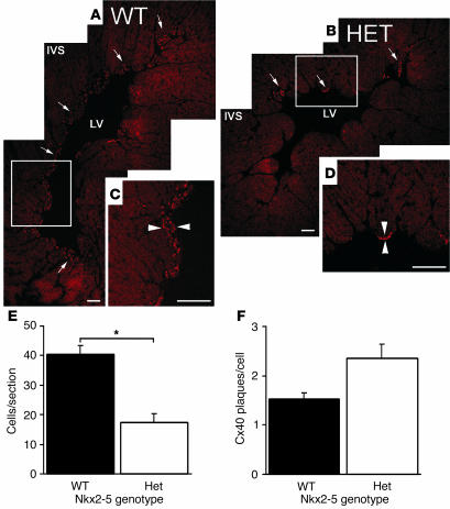
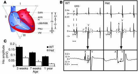
Heterozygous mutations of the cardiac transcription factor Nkx2-5 cause atrioventricular conduction defects in humans by unknown mechanisms. We show in KO mice that the number of cells in the cardiac conduction system is directly related to Nkx2-5 gene dosage. Null mutant embryos appear to lack the primordium of the atrioventricular node. In Nkx2-5 haploinsufficiency, the conduction system has half the normal number of cells. In addition, an entire population of connexin40(-)/connexin45(+) cells is missing in the atrioventricular node of Nkx2-5 heterozygous KO mice. Specific functional defects associated with Nkx2-5 loss of function can be attributed to hypoplastic development of the relevant structures in the conduction system. Surprisingly, the cellular expression of connexin40, the major gap junction isoform of Purkinje fibers and a putative Nkx2-5 target, is unaffected, consistent with normal conduction times through the His-Purkinje system measured in vivo. Postnatal conduction defects in Nkx2-5 mutation may result at least in part from a defect in the genetic program that governs the recruitment or retention of embryonic cardiac myocytes in the conduction system.


Figures

Figure 1
Figure 1
The absence of the AV node primordium in Nkx2-5–null mutant embryos. (A) Schematic diagram of the Nkx2-5 genomic structure, targeting construct, and Nkx2-5neo allele and Northern blot analysis demonstrating a 50% reduction in Nkx2-5 mRNA in the Nkx2-5+/neo adult ventricular myocardium. (B) WT Nkx2-5 E9.5 embryos that carry the minK-lacZ gene show blue X-gal staining in the inner curvature of the AV canal (arrow). (C) Nkx2-5neo/neo E9.5 embryos show abnormal development of the heart tube and no minK-lacZ activity in the AV canal region where the AV node primordium is expected (arrow); staining is normal in the somites (arrowhead). In situ hybridization for minK mRNA and the corresponding brightfield images demonstrate expression throughout the myocardium of WT (D and E) and Nkx2-5neo/neo (F and G) embryos. All images are representative of three or more embryos or mice. Comparisons of minK-lacZ expression were made between animals with identical copy numbers of the reporter allele. B and C show minK-lacZ homozygotes; heterozygotes yield similar results. WT, Nkx2-5 WT; Het, Nkx2-5+/neo; KO, Nkx2-5neo/neo; H, HindIII; N, NotI; S, SpeI; e 1, exon; e 2, exon 2; L, loxP sequence; neo, pGK neomycin resistance cassette. Probe denotes fragments used for Southern blot analysis. Scale bars: 200 μm (B and C), 100 μm (D–G).
Figure 2
Figure 2
Hypoplasia of the central conduction system in heterozygous Nkx2-5 KO mice. Histologic sections of adult hearts demonstrate the blue, X-gal–stained cells of the AV node (A and B) and His bundle (C and D) in WT (A and C) and Nkx2-5+/neo (B and D) hearts bearing one minK-lacZ allele. The histology of the AV node and His bundle were also examined by H&E and immunohistochemical staining of WT (E, F, I, J) and Nkx2-5+/– (G, H, K, L) hearts. Cx40 (TRITC, red) and Cx43 (FITC, green) label conductive and contractile myocytes, respectively. The AV node (F and H) and His bundle tissues (J and L) are circled. Consecutive sister sections from the same AV nodes were double-labeled for Cx40 (FITC, green) and Cx45 (TRITC, red) (circled in M and N). In the WT node (M), a small population of Cx40+/Cx45+ cells is present (arrow), but a larger population bearing Cx45 alone predominates. In the Nkx2-5+/– node (N) the Cx40+/Cx45+ cells are present, but the population expressing Cx45 alone is absent. Images shown are representative of at least three separate animals. HET, Nkx2-5+/– or Nkx2-5+/neo; AVN, AV node; His, His bundle. Scale bars: 100 μm.
Figure 3
Figure 3
Hypoplasia of the peripheral conduction system in Nkx2-5neo/+ mice revealed by minK-lacZ expression. (A) In E14.5 hearts minK-lacZ enzymatic activity is proportional to minK-lacZ (copy numbers 0, 1, or 2) and Nkx2-5 gene dosage. Whole-mount images of minK-lacZ–stained neonatal hearts contrast the dense Purkinje fiber network in WT (B and D) compared with the hypocellular system in Nkx2-5+/neo mice (C and E). Sections show that the prominent blue X-gal stain shown in the whole mount is the peripheral conduction system at the interventricular septum (D and E). The hearts shown were from minK-lacZ homozygotes; heterozygotes yield similar results. The images shown are representative of at least three hearts. P < 0.05.
Figure 4
Figure 4
Cx40 immunohistochemistry confirms hypoplasia of the peripheral Purkinje system and normal cellular expression levels of Cx40 in Nkx2-5+/– mice. Montage confocal images demonstrate Cx40 expression as red, punctate staining in the subendocardial Purkinje fibers of WT (A and C) and Nkx2-5+/– (B and D) left ventricular myocardium. The distribution of Cx40 in the Nkx2-5+/– myocardium is considerably smaller than in WT (arrows in A and B). Higher magnification images of the boxed areas in A and B reveal the increased thickness of Purkinje fiber layers (arrowheads) in the WT (C) compared with Nkx2-5+/– heart (D). LV, left ventricle. Scale bars: 100 μm. (E) Cell counts within Cx40-positive domains reveal that Purkinje cell numbers are reduced by approximately half within sections of Nkx2-5+/– ventricles (P < 0.05). (F) Nkx2-5+/– and WT Purkinje cells contain approximately the same the number of Cx40 particles per cell. Three hearts each from WT and Nkx2-5+/– animals were examined.
Figure 5
Figure 5
ECG correlates of anatomic structures in the cardiac conduction system in WT and Nkx2-5+/– mice. (A) Schematic diagram of the cardiac conduction system and ECG intervals as symbolized by the surface vectors I, II, and III and the intracardiac catheter. (B) Representative surface and intracardiac electrogram from a WT and Nkx2-5+/– mouse. The upper and middle tracings are simultaneous surface ECGs and IEGMs. The lower tracing shows the intracardiac recording at higher magnification and demonstrates the difference in His bundle signal amplitude between WT and Nkx2-5+/– mice. Atrial (P, A) and ventricular (QRS, V) depolarizations are depicted. The IEGM shows His (H) depolarization. Atrio-His and His-ventricular intervals are denoted (1 and 2). Note that ventricular depolarization or QRS interval begins immediately after the HV interval. (C) The amplitude of His depolarization is diminished in Nkx2-5+/– mice at all ages examined (P < 0.0001 in all age groups). AV, atrial-ventricular; AH, atrio-His; HV, His-ventricular; LBB/RBB, left and right bundle branch; PMJ, Purkinje-myocyte junction; SAN, sinoatrial node; VM, ventricular myocardium.

References

    1. Wang, G.F., and Stockdale, F.E. 1999. Chamber-specific gene expression and regulation during heart development. In Heart development. R.P. Harvey and N. Rosenthal, editors. Academic Press. San Diego, California, USA. 357–369.
    1. Cheng G, et al. Development of the cardiac conduction system involves recruitment within a multipotent cardiomyogenic lineage. Development. 1999;126:5041–5049. - PubMed
    1. Gourdie RG, Mima T, Thompson RP, Mikawa T. Terminal diversification of the myocyte lineage generates Purkinje fibers of the cardiac conduction system. Development. 1995;121:1423–1431. - PubMed
    1. Sedmera D, et al. Spatiotemporal pattern of commitment to slowed proliferation in the embryonic mouse heart indicates progressive differentiation of the cardiac conduction system. Anat. Rec. 2003;274A:773–777. - PubMed
    1. Gourdie RG, Wei Y, Kim D, Klatt SC, Mikawa T. Endothelin-induced conversion of embryonic heart muscle cells into impulse-conducting Purkinje fibers. Proc. Natl. Acad. Sci. U. S. A. 1998;95:6815–6818. - PMC - PubMed

Publication types