. 2004 Apr;113(8):1138-48.
doi: 10.1172/JCI19890.

Targeted deletion of BMK1/ERK5 in adult mice perturbs vascular integrity and leads to endothelial failure

Affiliations

Targeted deletion of BMK1/ERK5 in adult mice perturbs vascular integrity and leads to endothelial failure

Masaaki Hayashi et al. J Clin Invest. 2004 Apr.

Abstract

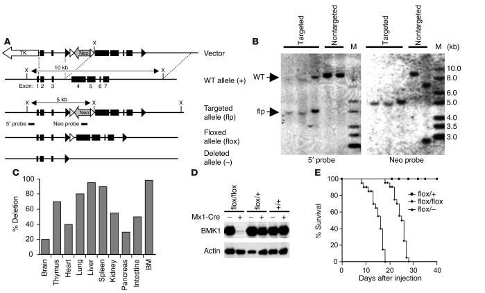
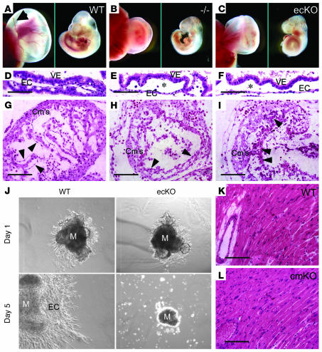
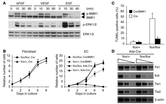
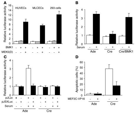
Big mitogen-activated protein kinase 1 (BMK1), also known as ERK5, is a member of the MAPK family. Genetic ablation of BMK1 in mice leads to embryonic lethality, precluding the exploration of pathophysiological roles of BMK1 in adult mice. We generated a BMK1 conditional mutation in mice in which disruption of the BMK1 gene is under the control of the inducible Mx1-Cre transgene. Ablation of BMK1 in adult mice led to lethality within 2-4 weeks after the induction of Cre recombinase. Physiological analysis showed that the blood vessels became abnormally leaky after deletion of the BMK1 gene. Histological analysis revealed that, after BMK1 ablation, hemorrhages occurred in multiple organs in which endothelial cells lining the blood vessels became round, irregularly aligned, and, eventually, apoptotic. In vitro removal of BMK1 protein also led to the death of endothelial cells partially due to the deregulation of transcriptional factor MEF2C, which is a direct substrate of BMK1. Additionally, endothelial-specific BMK1-KO leads to cardiovascular defects identical to that of global BMK1-KO mutants, whereas, surprisingly, mice lacking BMK1 in cardiomyocytes developed to term without any apparent defects. Taken together, the data provide direct genetic evidence that the BMK1 pathway is critical for endothelial function and for maintaining blood vessel integrity.


Figures

Figure 1
Figure 1
Conditional gene targeting of the BMK1 gene. (A) Diagram of the WT BMK1 genomic locus (+), the targeted BMK1 allele (flp), the floxed BMK1 allele (flox), and the BMK1 null allele (–). Open and closed arrowheads indicate FRT and loxP sites, respectively. (B) Southern blot analysis of targeted ES cell clones. DNAs were digested with XhoI and hybridized with external 5′ probe or internal Neo probe. M, marker. (C) Efficiency of Mx1-Cre–mediated BMK1 deletion in various organs. Hybridization signals from Southern blot analysis were quantified, and the deletion efficiency was calculated as the percentage of signals of deleted alleles in the total signals. BM, bone marrow. (D) Immunoblot analysis of BMK1 protein in the livers from pIpC-treated BMK1flox/flox, BMK1flox/+, and BMK1+/+ mice carrying or not carrying the Mx1-Cre transgene as indicated. (E) Mortality of BMK1-CKO mice after pIpC treatment: n = 12 for BMK1flox/+ mice; n = 20 for BMK1flox/flox mice; n = 20 for BMK1flox/– mice.
Figure 2
Figure 2
Efficiency of Cre-mediated recombination by induced Mx1-Cre transgene. (A) Schematic representation of the expression of Z/AP transgene before and after Cre recombination. (B) Sections from pIpC-treated double transgenic (Z/AP, Mx1-Cre) mice were stained for LacZ and/or AP activity. SMC, smooth muscle cell. Scale bars: 50 μm in brain, heart, and lung; 10 μm in aorta.
Figure 3
Figure 3
Increased vascular leakage after ablation of BMK1. (A–D) Representative ears showing increased baseline leakage of Evans blue dye in BMK1-CKO mice, which are further enhanced after topical application of mustard oil. Evans blue dye (30 mg/kg in a volume of 100 μl) was injected into the tail vein of anesthetized mice. (E) Quantification of extravasated Evans blue dye. Baseline leakage was elevated in BMK1-CKO mice (black bars) as compared with control WT mice (white bars). Similarly, significant increase in vascular leakage of BMK1-CKO mice was observed after mustard oil treatment. Values are mean ± SEM; n = 6 mice per group. *P < 0.05.
Figure 4
Figure 4
Histopathological analyses of pIpC-treated BMK1-CKO mice. (A) Histological analysis of BMK1-CKO hearts and pulmonary arteries at various time points after pIpC treatment. (B) Immunohistochemical analysis of control heart (left) and BMK1-CKO heart (right) with the CD31 Ab. (C) Immunofluorescent analysis of control heart (left) and BMK1-CKO heart (right) with Ab against smooth muscle actin (green) with nuclear DAPI staining (pseudo-colored, red). Arrowheads indicate abnormal endothelial nuclei. Asterisks indicate coronary arteries. Scale bars: A and B, 10 μm; C, 5 μm.
Figure 5
Figure 5
Analysis of apoptotic changes in BMK1-CKO mice. (A–D) Ultrastructure analyses of capillaries in BMK1-CKO mice. Transmission electron microscopy was performed in the capillaries of control heart (A) and BMK1-CKO heart (B−D). (E and F) TUNEL and CD31 immunohistochemical staining of heart sections. Confocal immunofluorescent analysis of heart sections stained with CD31 (red) and TUNEL (green) from control mice (E) and BMK1-CKO mice (F). (G) Quantitative analysis of EC apoptosis. Values are mean ± SEM. Asterisk indicates electron-lucent EC. Cm, cardiomyocyte; E, erythrocyte; L, lumen; P, pericyte. Arrow indicates the fenestration of the capillary. Scale bars: A−D, 2 μm; E and F, 10 μm.
Figure 6
Figure 6
Essential role of BMK1 in ECs but not in cardiomyocytes during embryogenesis. (A–C) Growth retardation of global BMK1-KO (BMK1–/–) and endothelial-specific BMK1-KO (BMK1-ecKO) embryos. Gross appearance of yolk sacs (left panel) and embryos (right panel) from WT (A), BMK1–/– (–/–) (B), and BMK1-ecKO (ecKO) (C) mutants at E9.5 are shown. Arrow in A indicates the major vitelline vessels, which are barely detectable in BMK1–/– and BMK1-ecKO mutants. (D–F) H&E staining of yolk sac of WT (D), BMK1–/– (E), and BMK1-ecKO (F) mutants at E9.5. Note that the vessels of the BMK1–/– and BMK1-ecKO yolk sacs are dilated compared with the vessels of the WT yolk sac. Asterisk denotes the underdeveloped vessels. (G–I) H&E staining of that hearts of WT (G), BMK1–/– (H), and BMK1-ecKO (I) mutants at E9.5. Arrowheads indicate ECs. (J) Deficit in endothelial proliferation in BMK1-ecKO heart explant cultures. (K and L) H&E staining of sections from WT and BMK1-cmKO (cmKO) hearts. M, myocardium; VE, visceral endoderm. Scale bars: D–I, K, and L, 10 μm.
Figure 7
Figure 7
Requirement of BMK1 for the survival of ECs. (A) Activation of BMK1 in ECs by angiogenic growth factors. After 24-hour serum starvation, ECs were stimulated with 10 ng/ml of growth factors for the interval indicated. p-BMK1, phospho-BMK1; p–ERK1/2, phospho–ERK1/2. (B) Growth curves of BMK1flox/+ and BMK1flox/flox fibroblast and EC cultures infected with either Ade-Cre or Ade-Cre along with Ade-BMK1 (Cre/BMK1). (C) Apoptosis in BMK1flox/+ and BMK1flox/flox EC cultures infected with Cre or Cre/BMK1 adenovirus scored by TUNEL assay. (D) Semiquantitative RT-PCR for VEGF and angiopoietin receptor. RNA samples are from BMK1flox/+ and BMK1flox/flox ECs infected with Cre-expressing adenovirus. A progression of fourfold dilutions of first-strand cDNA was used in each PCR to amplify gene products of Flt1, Kdr, Tie1, Tie2, and β-actin.
Figure 8
Figure 8
BMK1-mediated MEF2C activation is required for EC survival. (A) BMK1 stimulates MEF2C transactivation activity in ECs. HUVECs, MLCECs, and 293 cells were cotransfected with reporter plasmid pG5ElbLuc and GAL4-MEF2C. Additionally, control vector pcDNA3 or expression plasmids encoding MEK5(D) along with BMK1 were included in each transfection as indicated. (B and C) BMK1 mediates serum-induced MEF2C activation. MLCECs were first infected with adenovirus as indicated and were cotransfected with pG5E1bLuc and GAL4-MEF2C (B) or transfected with either pJLuc or pJSXLuc, cis-reporter plasmids of MEF2 element (C). Cells were then starved for 24 hours and stimulated with 10% serum for 6 hours before harvesting. (D) Expression of constitutively active MEF2C (MEF2C-VP16) rescued EC apoptosis caused by BMK1 deficiency. MLCECs were first infected with adenovirus as indicated and were cotransfected with GFP vector along with either control vector or MEF2C-VP16. Apoptosis in the transfected cell population (GFP-positive cells) was determined by cellular morphology 2 days after transfection. The data represent mean ± SEM of at least three independent transfections.

Comment in

References

    1. Kato Y, et al. BMK1/ERK5 regulates serum-induced early gene expression through transcription factor MEF2C. EMBO J. 1997;16:7054–7066. - PMC - PubMed
    1. Chao TH, Hayashi M, Tapping RI, Kato Y, Lee JD. MEKK3 directly regulates MEK5 activity as part of the big mitogen-activated protein kinase 1 (BMK1) signaling pathway. J. Biol. Chem. 1999;274:36035–36038. - PubMed
    1. English JM, Vanderbilt CA, Xu S, Marcus S, Cobb MH. Isolation of MEK5 and differential expression of alternatively spliced forms. J. Biol. Chem. 1995;270:28897–28902. - PubMed
    1. Zhou G, Bao ZQ, Dixon JE. Components of a new human protein kinase signal transduction pathway. J. Biol. Chem. 1995;270:12665–12669. - PubMed
    1. Kato Y, et al. Bmk1/Erk5 is required for cell proliferation induced by epidermal growth factor. Nature. 1998;395:713–716. - PubMed

Publication types

Substances