

Susceptibility of T cell receptor-alpha chain knock-out mice to ultraviolet B light and fluorouracil: a novel model for drug-induced cutaneous lupus erythematosus
- PMID: 15086387
- PMCID: PMC1809037
- DOI: 10.1111/j.1365-2249.2004.02458.x
Susceptibility of T cell receptor-alpha chain knock-out mice to ultraviolet B light and fluorouracil: a novel model for drug-induced cutaneous lupus erythematosus
Abstract
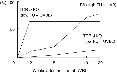
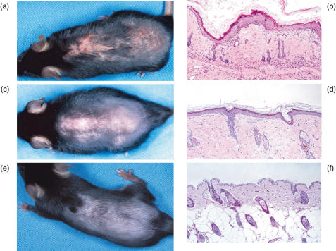
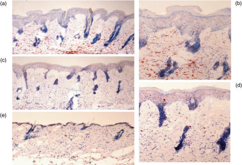
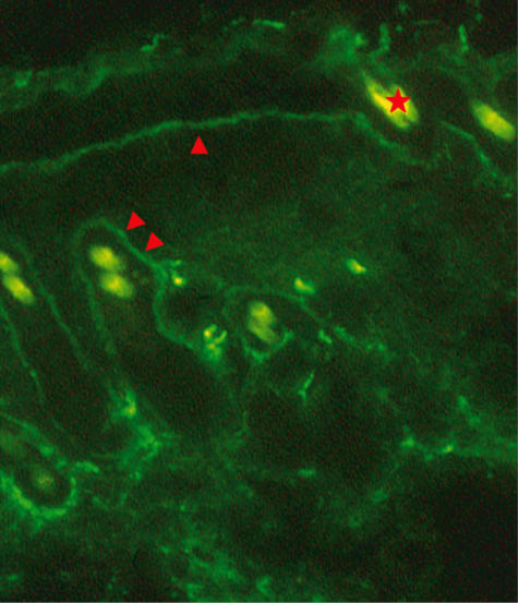
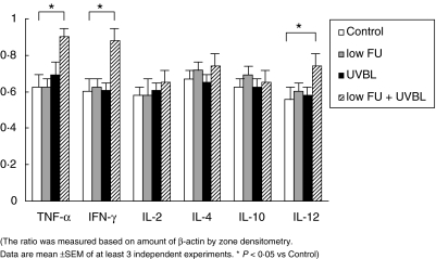
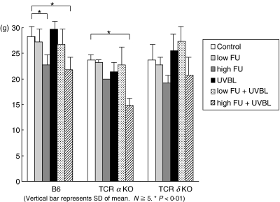
The anticancer agent 5-fluorouracil (FU) frequently induces cutaneous lupus erythematosus (LE) lesions on sun exposed sites. Based on this observation, we have tried to establish a cutaneous LE model of C57BL/6 J (B6) mice, B6 T cell receptor (TCR)-alpha(-/-) mice and B6 TCR-delta(-/-) mice treated with FU and/or ultraviolet B light (UVBL) in order to clarify the role of T cells and the cytokine profile of cutaneous lupus lesions. Cutaneous LE-like skin lesions could be induced in TCR-alpha(-/-) mice with low FU (0.2 mg) plus UVBL, and in B6 mice treated with a high dose of FU (2.0 mg) plus UVBL. In contrast, low FU plus UVBL induced such skin lesions in TCR-delta(-/-) mice at a very low incidence. Specifically, the skin lesions of TCR-alpha(-/-) mice with low FU plus UVBL appeared more rapidly and were more severe than lesions in B6 mice. The former had the common characteristic features of human chronic cutaneous LE such as typical histology, positive IgG at the dermoepidermal junction, low antinuclear antibody and low mortality. Furthermore, a Th1 response was induced in the development of drug-induced cutaneous LE. FU and UVBL-induced cutaneous LE-like eruption is an excellent model for better understanding the pathomechanisms of skin lesion development in LE.
Figures
Comment in
-
Drugs, sun and T cells in lupus.Clin Exp Immunol. 2004 May;136(2):191-3. doi: 10.1111/j.1365-2249.2004.02455.x. Clin Exp Immunol. 2004. PMID: 15086379 Free PMC article. No abstract available.
References
-
- Elder D, Elenitsas R, Jaworsky C, et al. Connective tissue diseases. In: Elder D, Elenitsas R, Jaworsky C, Johnson B Jr, editors. Lever's histopathology of the skin. 8. Philadelphia: Lippincott-Raven Publishers; 1997. pp. 253–85.
-
- Norris DA. Pathomechanisms of photosensitive lupus erythematosus. J Invest Dermatol. 1993;100:58s–68s. - PubMed
-
- Hasen T, Nyberg F, Stephansson E, et al. Photosensitivity in lupus erythematosus, UV photoprovocation results compared with history of photosensitivity and clinical findings. Br J Dermatol. 1997;136:699–705. - PubMed
-
- Stein LF, Saed GM, Fivenson DP. T-cell cytokine network in cutaneous lupus erythematosus. J Am Acad Dermatol. 1997;36:191–6. - PubMed
-
- Hoffman BJ. Sensitivity to sulfadazine resembling acute disseminated lupus erythematosus. Arch Dermatol Syph. 1945;51:190–2.
Publication types
MeSH terms
Substances
LinkOut - more resources
Full Text Sources
Molecular Biology Databases
