. 2004 Jun;93(6):741-53.
doi: 10.1093/aob/mch092. Epub 2004 Apr 15.

Calcium channels are involved in calcium oxalate crystal formation in specialized cells of Pistia stratiotes L

Affiliations

Calcium channels are involved in calcium oxalate crystal formation in specialized cells of Pistia stratiotes L

Gayle M Volk et al. Ann Bot. 2004 Jun.

Abstract

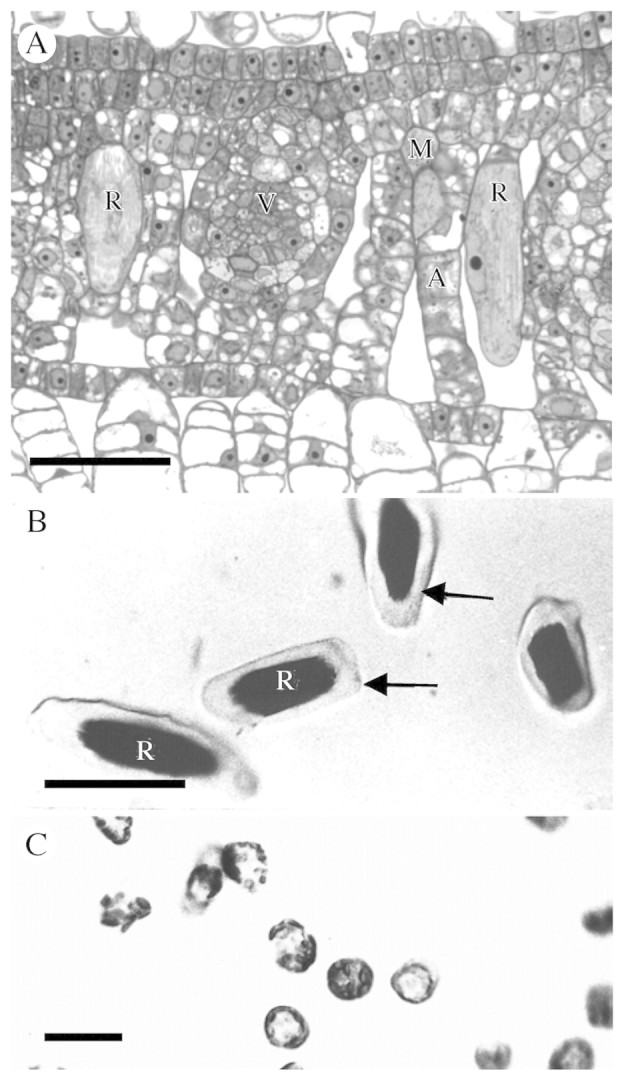
Background and aims: Pistia stratiotes produces large amounts of calcium (Ca) oxalate crystals in specialized cells called crystal idioblasts. The potential involvement of Ca(2+) channels in Ca oxalate crystal formation by crystal idioblasts was investigated.

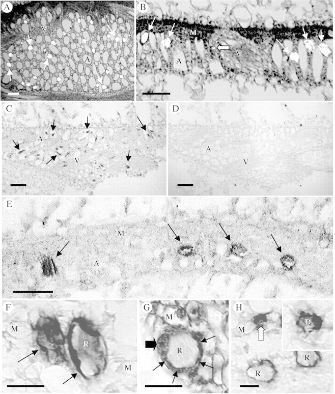
Methods: Anatomical, ultrastructural and physiological analyses were used on plants, fresh or fixed tissues, or protoplasts. Ca(2+) uptake by protoplasts was measured with (45)Ca(2+), and the effect of Ca(2+) channel blockers studied in intact plants. Labelled Ca(2+) channel blockers and a channel protein antibody were used to determine if Ca(2+) channels were associated with crystal idioblasts.

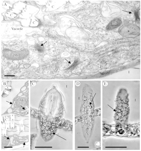
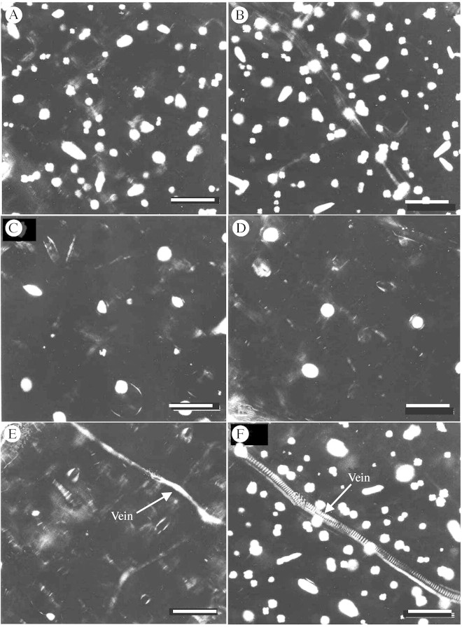
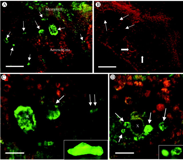
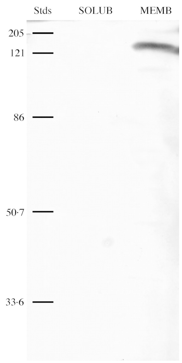
Key results: (45)Ca(2+) uptake was more than two orders of magnitude greater for crystal idioblast protoplasts than mesophyll protoplasts, and idioblast number increased when medium Ca was increased. Plants grown on media containing 1-50 microM of the Ca(2+) channel blockers, isradipine, nifedipine or fluspirilene, showed almost complete inhibition of crystal formation. When fresh tissue sections were treated with the fluorescent dihydropyridine-type Ca(2+) channel blocker, DM-Bodipy-DHP, crystal idioblasts were intensely labelled compared with surrounding mesophyll, and the label appeared to be associated with the plasma membrane and the endoplasmic reticulum, which is shown to be abundant in idioblasts. An antibody to a mammalian Ca(2+) channel alpha1 subunit recognized a single band in a microsomal protein fraction but not soluble protein fraction on western blots, and it selectively and heavily labelled developing crystal idioblasts in tissue sections.

Conclusions: The results demonstrate that Ca oxalate crystal idioblasts are enriched, relative to mesophyll cells, in dihydropyridine-type Ca(2+) channels and that the activity of these channels is important to transport and accumulation of Ca(2+) required for crystal formation.


Figures

None
Fig. 1. Raphide idioblasts and protoplasts of Pistia stratiotes. (A) Cross‐section of young leaf containing two developing raphide idioblasts, which are much larger than surrounding cells. (B) Isolated raphide idioblast protoplasts (arrows). (C) Mesophyll protoplasts. Scale bars: A = 40 µm; B = 50 µm; C = 20 µm. A, Aerenchyma; M, mesophyll; R, raphide crystal idioblast; V, vein.
None
Fig. 2. Endoplasmic reticulum and cytoplasmic features of raphide crystal idioblasts. (A and B) Transmission electron micrographs of part of a developing raphide idioblast cytoplasm and vacuole. (A) Smooth and rough ER, Golgi (arrows) and other membrane structures are abundant in the cytoplasm. (B) Part of a developing idioblast showing regions of enlarged ER (arrows) and the membranes associated with the edges of the crystals in the vacuole. (C–E) Phase contrast images of paraffin sections through raphide idioblasts. (C) Tangential section through an idioblast. The crystal bundle is visible at the top of the cell and the reticulate peripheral cytoplasm (arrow) visible at the bottom. (D) Longitudinal section along the peripheral cytoplasm of an entire idioblast. A reticulate network can be seen (arrows). (E) Enlarged section along the peripheral cytoplasm of an idioblast, showing dense reticulate pattern. Scale bars: A and B = 1 µm; C–E = 20 µm. A, Aerenchyma; C, crystal; I, intercellular space; Mt, mitochondrion; P, plastid; RER, rough endoplasmic reticulum, W, wall.
None
Fig. 3. Effect of Ca2+ channel blockers on Ca oxalate formation. Images are of representative clearings from whole leaves that developed on plants during growth on media containing blocker or control agents. Crystal idioblasts appear as bright spots in these images viewed between crossed polarizing filters. (A) Leaf from plant on normal growth medium with 2·5 mm Ca. (B) Leaf from plant on medium supplemented with 5 mm Ca. There is about a 25 % increase in crystal idioblasts, demonstrating that crystal amount is related to Ca availability. (C) Leaf from plant treated with 1 µm fluspirilene in 0·5 % DMSO. Crystal formation is greatly inhibited. (D) Leaf from plant treated with 50 µm isradipine. Crystal formation is almost completely inhibited. (E) Leaf from plant treated with 50 µm nifedipine. Crystal formation is completely inhibited. (F) Leaf from plant on 0·5 % DMSO. There is no inhibition of crystal formation. Scale bars = 400 µm.
None
Fig. 4. Binding of green fluorescent‐tagged Ca2+ channel blocker, DM‐Bodipy‐DHP, to fresh sections of developing Pistia leaf. (A) Low magnification image. The brightest green fluorescence is associated with Ca oxalate crystal idioblasts at various stages of development (arrows). Some fluorescence is also seen in the young developing mesophyll cells. Red fluorescence is due to chlorophyll. (B) Control. Binding of DM‐Bodipy‐DHP is completely inhibited by a 4‐h pretreatment with the competitive Ca2+ channel blocker, nifedipine. Raphide crystal idioblasts (arrows) and druse idioblasts (block arrows) are pointed out for reference. (C) Enlargement from A showing labelled crystal idioblasts. The raphide idioblast to the left (R) is focused through the cortical cytoplasm indicating Ca2+ channels are abundant in cytoplasmic organelles. At the large single arrow, the same focal plane is near the surface of the raphide idioblast indicating potential association of the fluorescent probe with the plasma membrane as well. At the double arrow is a cell that is just beginning initiation of differentiation to become a raphide idioblast. Inset is a living raphide crystal idioblast stained with DiOC6 to show ER abundance. (D) Image of the xylem region of a vein where druse crystal idioblasts are most common. At least four druse idioblasts at various developmental stages are seen and are fluorescing brightly (arrows). Inset is of living druse idioblasts stained with DiOC6. R, Raphide crystal idioblast; X, xylem. Scale bars: A and B = 50 µm; C and D = 21 µm.
None
Fig. 5. Western immunoblot of soluble protein (SOLUB) and microsomal membrane protein (MEMB) from Pistia extracts (100 µg protein loaded for each). The blot was probed with an antibody (MAB427) to an α1 subunit of a DHP‐sensitive Ca2+ channel. For reference, relative molecular weight standards (Stds) are given in kDa. A single band of about 175 kDa was detected in the membrane but not soluble fraction.
None
Fig. 6. Immunological localization of putative Ca2+ channels (α1 subunit of a DHP‐sensitive Ca channel) in paraffin sections of developing Pistia leaf tissue. Immunolabelled sections were not counterstained. (A, B and G) Sections of developing leaves stained for general histology for reference. (A) Paradermal section through aerenchyma, viewed with partially crossed polarizing filters. Crystal idioblasts (bright spots, arrows) are distributed throughout the aerenchyma. (B) Cross‐section showing developing raphide idioblasts (arrows) and a druse idioblast (block arrow). (C) Tangential section mostly through aerenchyma of a developing leaf that has been immunolabelled. The many developing idioblasts (arrows) are strongly stained, appearing as dark spots relative to the surrounding cell types. (D) Serial section to that in C but treated only with the secondary antibody. There is no endogenous peroxidase activity or nonspecific binding of the secondary antibody in the paraffin‐embedded sections. The idioblasts are not apparent. (E) Cross‐section through a developing leaf immunostained for α1 subunit of a DHP‐sensitive Ca2+ channel. Four developing idioblasts can be seen (arrows) and are heavily stained. The idioblast on the left is sectioned along the surface and appears completely dark, while the three idioblasts to the right show staining primarily along the periphery and not in the vacuole. (F) Two raphide idibolasts, one sectioned along the surface and one as a cross‐section. The label can be seen associated with the preipheral region. (G) General staining of a developing raphide idioblast for comparison to immunolabel, showing dense ring of cytoplasm (arrows) surrounding the crystal bundle. The enlarged nucleus (big arrow) can also be seen. (H) Section showing two raphide idioblasts and a druse idioblast (block arrow). The idioblasts label strongly with the antibody, but mesophyll cells show no significant label. Inset: Druse idioblast where the star‐shaped druse crystal can be seen surrounded by dense staining material from the cortical region of the cell. Scale bars: A–E = 100 µm, F–G = 20 µm. A, aerenchyma; D, druse crystal; M, mesophyll; R, raphide crystal bundle; V, vein.

References

    1. AllenGJ, Sanders D.1994. Two voltage‐gated, calcium release channels coreside in the vacuolar membrane of broad bean guard cells. Plant Cell 6: 685–694. - PMC - PubMed
    1. AndrejauskasE, Hertel R, Marme D.1985. Specific binding of the calcium antagonist [3H]verapamil to membrane fractions from plants. Journal of Biological Chemistry 260: 5411–5414. - PubMed
    1. ArnottHJ.1982. Three systems of biomineralization in plants with comments on the associated organic matrix. In: Nancollas GH, ed. Biological mineralization and demineralization. Berlin: Springer‐Verlag, 199–218.
    1. ArnottHJ, Pautard FGE.1970. Calcification in plants. In: Schraer H, ed. Biological calcification. Cellular and molecular aspects. New York: Appleton‐Century‐Crofts, 375–446.
    1. BhatlaSC, Kiessling J, Reski R.2002. Observation of polarity induction by cytochemical localization of phenylalkylamine‐binding sites in regenerating protoplasts of the moss Physcomitrella patens Protoplasma 219: 99–105. - PubMed

Publication types

MeSH terms