

doi: 10.1042/BJ20040523.
Segregation of two glutaminase isoforms in islets of Langerhans
Affiliations
- PMID: 15089745
- PMCID: PMC1133855
- DOI: 10.1042/BJ20040523
Item in Clipboard
Segregation of two glutaminase isoforms in islets of Langerhans
Biochem J.
.
Abstract
Despite the importance of glutamatergic signalling in the co-ordination of hormone secretion, the identity of the enzyme for the production of glutamate in beta-cells is still unresolved. We have found that the endocrine pancreas co-expresses two isoforms of GA (glutaminase), denoted as kidney-type (KGA) and liver-type (LGA), with a complementary cellular pattern of expression. Whereas KGA was mainly present in alpha-cells, LGA was very abundant in beta-cells. This spatial segregation may have important functional implications, facilitating a differential regulation of glutamate production in insulin- and glucagon-secreting cells.
Figures

Representative Western blots showing the presence of both GA isoforms (LGA and KGA) in islets isolated from rat pancreas. To assess potential cross-reactivity and to illustrate the isoform specificity of our purified anti-GA antibodies, rat brain and liver samples were immunoblotted as described in the text.
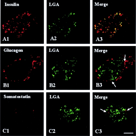
Double-immunofluorescence labelling with anti-LGA (green) and endocrine cell markers: insulin, glucagon or somatostatin (red). In confocal overlay images, yellow represents co-localization of the two antigens. (A3) LGA and insulin, (B3) LGA and glucagon and (C3) LGA and somatostatin. Note that, whereas most of the insulin-containing cells exhibited a strong LGA immunoreactivity, only a few α- and δ-cells showed a weak, albeit detectable (arrows), immunolabelling. Scale bar, 57 μm.
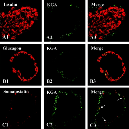
Double-immunofluorescence labelling with anti-KGA (green) and endocrine cell markers: insulin, glucagon or somatostatin (red). In confocal overlay images, yellow represents co-localization of the two antigens. (A3) KGA and insulin, (B3) KGA and glucagon and (C3) KGA and somatostatin. KGA was present in α- and δ-cells (B3 and C3 respectively) but was not detected in insulin-containing cells (A3). Scale bar, 57 μm.
References
-
- Moriyama Y., Hayashi M. Glutamate-mediated signalling in the islets of Langerhans: a thread entangled. Trends Pharmacol. Sci. 2003;24:511–517. - PubMed
-
- Maechler P., Wollheim C. B. Mitochondrial glutamate acts as a messenger in glucose-induced insulin exocytosis. Nature (London) 1999;402:685–689. - PubMed
-
- Nicklas W. J., Zeevalk G., Hyndman A. Interactions between neurons and glia in glutamate/glutamine compartmentation. Biochem. Soc. Trans. 1987;15:208–210. - PubMed
-
- Inagaki N., Kuromi H., Gonoi T., Okamoto Y., Ishida H., Seino Y., Kaneko T., Iwanaga T., Seino S. Expression and role of ionotropic glutamate receptors in pancreatic islet cells. FASEB J. 1995;9:686–691. - PubMed
-
- Hoy M., Maechler P., Efanov A. M., Wollheim C. B., Berggren P. O., Gromada J. Increase in cellular glutamate levels stimulates exocytosis in pancreatic β-cells. FEBS Lett. 2002;531:199–203. - PubMed
Publication types
MeSH terms
Substances
LinkOut - more resources
Full Text Sources