

Case Reports
A rare case of a ruptured middle meningeal aneurysm causing intracerebral hematoma in a patient with moyamoya disease
Affiliations
- PMID: 15090344
- PMCID: PMC7975590
Item in Clipboard
Case Reports
A rare case of a ruptured middle meningeal aneurysm causing intracerebral hematoma in a patient with moyamoya disease
AJNR Am J Neuroradiol.
2004 Apr.
Abstract
Moyamoya disease is infrequently associated with intracranial aneurysms arising from the circle of Willis vessels or from "peripheral" branches of choroidal and meningeal vessels. We present a rare case of a moyamoya-related aneurysm arising along the dural junction of multiple meningeal branches from the external carotid artery causing intracerebral hemorrhage. Endovascular coil embolization of the middle meningeal artery (MMA) and occipital artery (OA) led to delayed aneurysm obliteration without rehemorrhage.
Figures
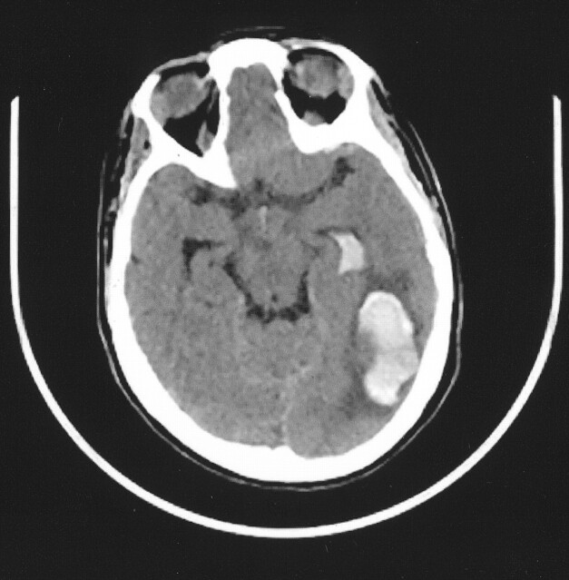
CT scan, showing left occipito-parietal hematoma with intraventricular hemorrhage.
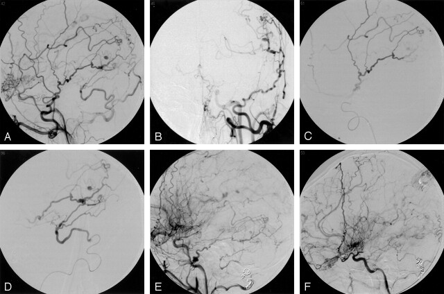
A and B, Left common carotid artery lateral and AP arteriogram, demonstrating a 3-mm aneurysm arising near collateral branches of the MMA and OA. C, Superselective arteriogram of the MMA, demonstrating supply to the aneurysm D, Superselective arteriogram of the OA, demonstrating supply to the aneurysm. E, Left external carotid arteriogram, lateral view, demonstrating coil deposition in the MMA and OA with delayed aneurysm filling from a small external carotid artery branch unable to be catheterized. F, Two-month follow-up left external carotid arteriogram, showing complete obliteration of the aneurysm.
References
-
- Borota L, Marinkovic S, Kovacevic M. Intracranial aneurysms associated with moyamoya disease. Neurol Med Chir (Tokyo) 1996;36:860–864 - PubMed
-
- Kawagushi S, Sakaki T, Morimoto T, et al. Characteristics of intracranial aneurysms associated with moyamoya disease: a review of 111 cases. Acta Neurochir (Wien) 1996;138:1287–1294 - PubMed
-
- Yoshida Y, Yoshimoto T, Shirane R, Sakurai Y. Clinical course, surgical management, and long-term outcome of moyamoya patients with rebleeding after an episode of intracerebral hemorrhage. Stroke 1999;30:2272–2276 - PubMed
-
- Hamada J-I, Hashimoto N, Tsukahara T. Moyamoya disease with repeated intraventricular hemorrhage due to aneurysm rupture. J Neurosurg 1994;80:328–331 - PubMed
-
- Kuroda S, Houkin K, Kamiyama H, Abe H. Effects of surgical revascularization on peripheral artery aneurysms in moyamoya disease: report of three cases. Neurosurgery 2001;49:463–468 - PubMed
Publication types
MeSH terms
LinkOut - more resources
Full Text Sources
Medical