

Case Reports
Erdheim-Chester disease: MR imaging, anatomic, and histopathologic correlation of orbital involvement
Affiliations
- PMID: 15090356
- PMCID: PMC7975611
Item in Clipboard
Case Reports
Erdheim-Chester disease: MR imaging, anatomic, and histopathologic correlation of orbital involvement
AJNR Am J Neuroradiol.
2004 Apr.
Abstract
Erdheim-Chester disease (ECD) is a rare form of histiocytosis of unknown origin characterized by tissue infiltration by lipid-laden histiocytes. Typically, the diaphyseal and metaphyseal portions of the tubular bones are affected, leading to a characteristic radiographic pattern of bone sclerosis. Orbital involvement is not infrequent and is manifested by exophthalmos and periorbital xanthomatous lesions, with associated visual problems. This case report documents imaging and pathologic findings in a patient with ECD with extensive orbital involvement.
Figures
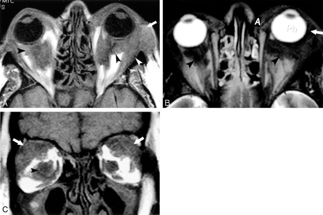
Images acquired 3 months before patient died. A, Axial T1-weighted image (TR/TE, 600/14; field of view [FOV], 14; section thickness, 3 mm skip 1 mm) demonstrating bilateral low-signal-intensity lesions in the intraconal (black arrowhead) and extraconal (white arrowhead) spaces, and anterior to the orbital septum (white arrow). B, Axial T2-weighted image (TR/TE, 3200/100; FOV, 14; section thickness, 3 mm skip 1 mm) showing similar low-signal-intensity lesions in the intraconal space (black arrowheads) and anterior to the orbital septum (white arrow). C, Coronal T1-weighted image (TR/TE, 600/14; FOV, −14; section thickness, 3 mm skip 1 mm) confirming low-signal-intensity lesions in the intraconal space (black arrowhead) and involvement of the lacrimal gland (white arrows).
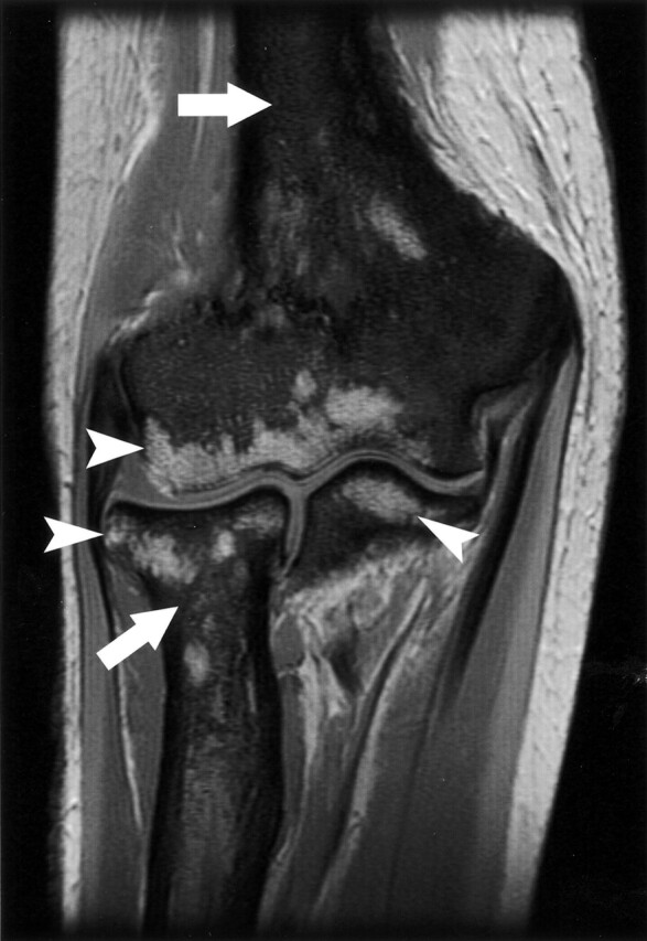
Coronal T1-weighted image of the elbow demonstrating the replacement of normal bone marrow by low-signal-intensity, diffuse lesions (white arrows) in the humerus, radius, and ulna. High-signal-intensity, normal fatty marrow is spared in portions of the epiphysis of those bones (white arrowheads).
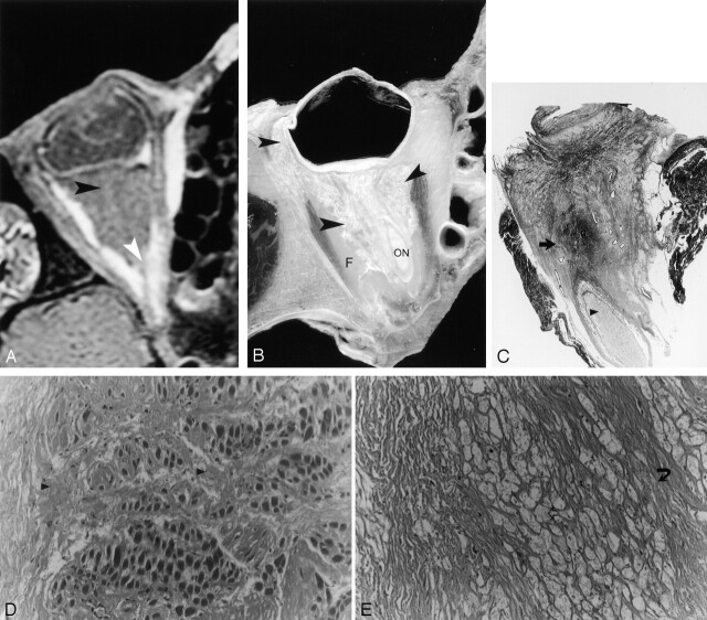
Postmortem correlation with MR imaging, cadaveric sections, and histology. A, Axial T1-weighted MR imaging (TR/TE, 600/20; FOV, 10; matrix, 256 × 196; section thickness, 3 mm skip 1 mm) showing low-signal-intensity lesions of the right orbit in the intraconal space (black arrowhead). The normal retro-orbital fat is replaced by abnormal diseased tissue and the optic nerve is encased (white arrowhead). The extraocular muscles do not show any apparent alteration. B, Cadaveric section showing the infiltrative disease (black arrowhead) at locations corresponding to the locations found with MR imaging. F+, retro-orbital fat; ON, optic nerve. C, Macropathology demonstrating abnormal replacement of the retro-orbital fat by a dense fibrous material and conglomerates of lipid-laden macrophages (black arrow), which encase the optic nerve (black arrowhead). Hematoxylin and eosin preparation. D, Histology with high magnification (magnification ×200) showing involvement of the muscular interstitial space by attenuated fibrous material (black arrowhead). E, Histology with high magnification (magnification ×100) demonstrating the characteristic lipid-laden macrophages of ECD (open arrow).
References
-
- Veyssier-Belot C, Cacoub P, Caparros-Lefebvre D, et al. Erdheim-Chester disease: clinical and radiological characteristics of 59 cases. Medicine (Balt) 1996;75:157–169 - PubMed
-
- Chester W. Uber Lipoidgranulomatose. Virchows Archiv 1930;279:561–602
-
- Jaffe HL. Metabolic, Degenerative and Inflammatory Diseases of Bones and Joints. Munich: Urban and Schwarzenberg;1970. :531–541
Publication types
MeSH terms
LinkOut - more resources
Full Text Sources
Medical