Localised invasive sino-orbital aspergillosis: characteristic features
- PMID: 15090423
- PMCID: PMC1772124
- DOI: 10.1136/bjo.2003.021725
Localised invasive sino-orbital aspergillosis: characteristic features
Abstract
Background/aim: To describe the characteristic constellation of historical, clinical, radiographic, and histopathological findings of localised invasive sino-orbital aspergillosis based on the authors' recent experience of four consecutive cases presenting over a 6 month period. Treatment and outcome are reviewed.
Methods: A case series of four patients with review of the English language literature.
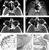
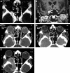
Results: There have been 17 reported cases of invasive sino-orbital aspergillosis in healthy individuals over the past 33 years. The authors report four patients who presented during a 6 month period with persistent and significant pain followed by progressive ophthalmic signs-clinical histories reflecting the literature. Similar imaging findings were also noted: focal hypodense areas within apical infiltrates on contrasted computed tomography correspond to abscesses seen at surgery, and sinus obliteration or involvement of the adjacent sinus lining was noted on magnetic resonance imaging. Bone erosion (often focal) was also seen. There is frequently a delay in making the correct diagnosis, and often disease progression occurs despite treatment.
Conclusions: The authors encountered four cases of invasive sino-orbital aspergillosis, three of which occurred in otherwise healthy individuals. The clinician must be aware of the characteristic presentation so that earlier diagnosis, management, and improved outcomes can be achieved.
Figures


References
-
- Levin LA, Avery R, Shore J, et al. The spectrum of orbital aspergillosis: a clinicopathological review. Surv Ophthalmol 1996;41:142–54. - PubMed
-
- Austin P, Dekker A, Kennerdell JS. Orbital aspergillosis. Report of a case diagnosed by fine needle aspiration biopsy. Acta Cytol 1983;27:166–9. - PubMed
-
- Bradley SF, McGuire NM, Kauffman CA. Sino-orbital and cerebral aspergillosis: cure with medical therapy. Mykosen 1987;30:379–85. - PubMed
-
- Fuchs HA, Evans RM, Gregg CR. Invasive aspergillosis of the sphenoid sinus manifested as a pituitary tumor. South Med J 1985;78:1365–7. - PubMed
-
- Green WR, Font RL, Zimmerman LE. Aspergillosis of the orbit. Arch Ophthalmol 1969;82:302–13. - PubMed
Publication types
MeSH terms
LinkOut - more resources
Full Text Sources
Medical
Miscellaneous
