. 2004 May;186(9):2655-63.
doi: 10.1128/JB.186.9.2655-2663.2004.

The membrane fluidity sensor DesK of Bacillus subtilis controls the signal decay of its cognate response regulator

Affiliations

The membrane fluidity sensor DesK of Bacillus subtilis controls the signal decay of its cognate response regulator

Daniela Albanesi et al. J Bacteriol. 2004 May.

Abstract

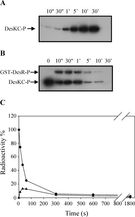
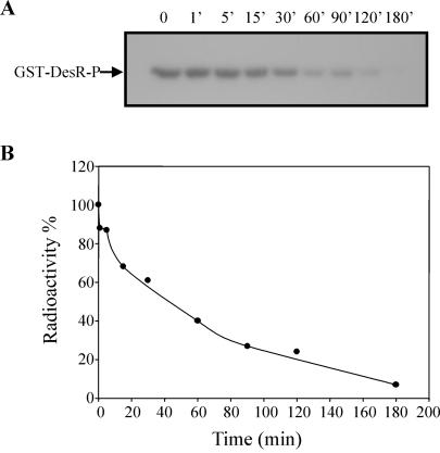
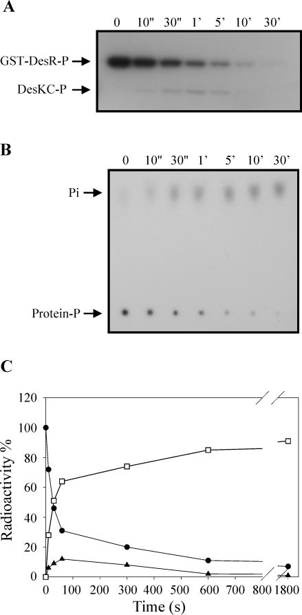
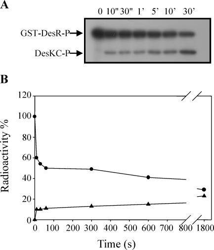
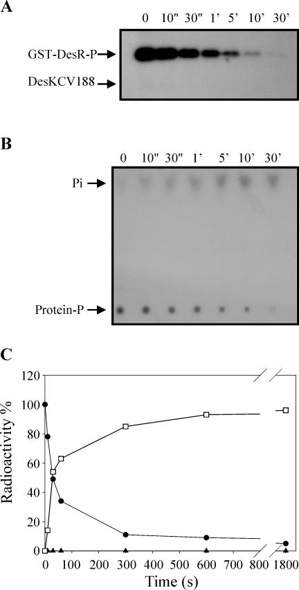
The Bacillus subtilis DesK/DesR two-component system regulates the expression of the des gene coding for the Delta5 acyl lipid desaturase. It is believed that a decrease in membrane lipid fluidity activates the DesK/DesR signal transduction cascade, which results in synthesis of the Delta5 acyl lipid desaturase and desaturation of membrane phospholipids. These newly synthesized unsaturated fatty acids then act as negative signals of des transcription, thus generating a regulatory metabolic loop that optimizes membrane fluidity. We previously suggested that DesK is a bifunctional enzyme with both kinase and phosphatase activities that could assume different signaling states in response to changes in the fluidity of membrane lipids. However, no direct experimental evidence supported this proposed model. In this study, we show that the C-terminal fragment of the DesK protein (DesKC) indeed acts as an autokinase. Addition of the response regulator DesR to phosphorylated DesKC resulted in rapid transfer of the phosphoryl group to DesR. Further, phosphorylated DesR can be dephosphorylated in the presence of DesKC, thus demonstrating that the sensor kinase has the ability to covalently modify DesR through both kinase and phosphatase activities. We also present evidence that DesKC might be locked in a kinase-dominant state in vivo and that its activities are not affected either in vivo or in vitro by unsaturated fatty acids. These findings provide the first direct evidence that the transmembrane segments of DesK are essential to sense changes in membrane fluidity and for regulating the ratio of kinase to phosphatase activities of the cytoplasmic C-terminal domain.


Figures

FIG. 1.
FIG. 1.
DesKC autophosphorylation and phosphotransfer from DesKC∼P to GST-DesR. (A) Purified DesKC, at a final concentration of 10 μM, was incubated in R buffer containing 25 μM ATP in the presence of 0.25 μCi of [γ-32P]ATP/μl at 25°C. The reaction was initiated by the addition of DesKC, and aliquots were removed at the indicated times. The reactions were stopped by the addition of 5× SDS gel loading buffer plus 10 mM EDTA and kept on ice until the last portion was taken. The samples were then subjected to SDS-12% PAGE analysis. After electrophoresis, the gel was dried and exposed for autoradiography. (B) DesKC was first allowed to autophosphorylate for 10 min as indicated in Materials and Methods, and then an equimolar fraction of GST-DesR was added to the reaction mixture. The reaction mixture was kept at 25°C, and aliquots were removed at the indicated times. The reactions were stopped and processed as described for panel A. (C) To quantitate the phosphoproteins, bands were cut from the gel and the incorporation of 32P was determined on a liquid scintillation counter. The label on DesKC∼P at the initiation of the reaction (0 s) was considered 100%. Circles correspond to DesKC∼P, and triangles correspond to GST-DesR∼P.
FIG. 2.
FIG. 2.
Stability of GST-DesR∼P. (A) GST-DesR∼P was purified as described in Materials and Methods and kept at 25°C in R buffer. Aliquots were removed at the indicated times, and the reactions were stopped by the addition of 5× SDS gel loading buffer plus 10 mM EDTA and processed as described in the legend to Fig. 1A. (B) The label on GST-DesR∼P at assayed times was determined as described in the legend to Fig. 1C. The label on GST-DesR∼P at the initiation of the experiment (0 min) was considered 100%. The ratio of remaining GST-DesR∼P was plotted versus time.
FIG. 3.
FIG. 3.
DesKC possesses GST-DesR∼P phosphatase activity. (A) GST-DesR∼P was purified as described in Materials and Methods in 150 μl of R buffer and mixed with 150 μl of equimolar DesKC in R buffer at 25°C. Aliquots were removed at the indicated times, and the reactions were stopped by the addition of 10× stop solution (10% SDS, 500 mM EDTA). Five percent of each time point sample was separated for TLC analysis (see below), and the rest was processed and subjected to SDS-PAGE as described in the legend to Fig. 1A. (B) TLC analysis was performed to analyze the destination of the label at each time point (see Material and Methods). Five percent of the samples was applied to a polyethyleneimine-cellulose plate (J. T. Baker) which was developed in 0.8 M LiCl and 0.8 M acetic acid. The plate was air dried and exposed to autoradiography films. (C) Quantitation of phosphoproteins from the gel was performed as described in the legend to Fig. 1C. To determine the ratio of released Pi to total phosphoproteins, spots from each time point were cut from the TLC plate and the radioactivity was determined on a liquid scintillation counter. The label on GST-DesR∼P at the initiation of the reaction (0 s) was considered 100%. Circles correspond to GST-DesR∼P, triangles correspond to DesKC∼P, and squares correspond to free Pi.
FIG. 4.
FIG. 4.
Effect of magnesium depletion on DesKC phosphatase activity. GST-DesR∼P was purified as described in Materials and Methods and mixed with an equimolar fraction of DesKC in R buffer at 25°C in the presence of 10 mM EDTA. Aliquots were removed at the indicated times, and the reactions were stopped and processed as described in the legend to Fig. 1A. (B) Quantitation of radioactivity on phosphoproteins was performed as described in the legend to Fig. 1C. The label on GST-DesR∼P at the initiation of the reaction (0 s) was considered 100%. Circles correspond to GST-DesR∼P, and triangles correspond to DesKC∼P.
FIG. 5.
FIG. 5.
His188 is not essential for DesKC phosphatase activity. Purified GST-DesR∼P in 150 μl of R buffer was mixed with 150 μl of equimolar DesKCV188 in R buffer at 25°C. Aliquots were removed at the indicated times, and reactions were stopped and processed as described in the legend to Fig. 3A. (B) TLC analysis was performed as described in the legend to Fig. 3B to analyze the destination of the label. (C) Quantitation of phosphoproteins was performed as described in the legend to Fig. 1C. Determination of the ratio of released Pi to phosphoproteins was perform as described in the legend to Fig. 3C. The label on GST-DesR∼P at the initiation of the reaction (0 s) was considered 100%. Circles correspond to GST-DesR∼P, triangles correspond to DesKCV188 (no DesKCV188∼P was detected), and squares correspond to free Pi.
FIG. 6.
FIG. 6.
His188 is not essential for DesKC phosphatase activity in vivo. Strains DA2095 (AKP20 thrC::pDG795), DA20K (AKP20 thrC::Pxyl-desK), and DA20KV (AKP20 thrC::Pxyl-desKV188) (Table 1) were grown overnight at 37°C in SMM supplemented with 0.01% threonine and 0.05% casein hydrolysate. Cells were collected and diluted either in the presence (grey bars) or in the absence (black bars) of 0.8% xylose. β-Galactosidase activities were determined 4 h after dilution. The results shown are the averages of the results from three independent experiments. UM, Miller units.
FIG. 7.
FIG. 7.
Expression of DesKC in vivo leads to constitutive transcription of des. Strain CM21 (Pdes-lacZ desKR mutant thrC::Pxyl-desR) (triangles) and strain CM21 transformed with plasmid pCM9 (Pxyl-desKC) (circles) were grown overnight in SMM supplemented with 0.01% threonine and 0.05% casein hydrolysate at 37°C. Cells were collected and diluted in the same medium in the presence (filled symbols) or absence (empty symbols) of 0.8% xylose. Samples were taken at 1-h intervals after dilution and assayed for β-galactosidase activity. Triangles superimpose with empty circles. Each datum point is the mean of the results from three independent experiments with a mean error of less than 5%. UM, Miller units.

References

    1. Aguilar, P. S., A. M. Hernandez-Arriaga, L. E. Cybulski, A. C. Erazo, and D. de Mendoza. 2001. Molecular basis of thermosensing: a two-component signal transduction thermometer in Bacillus subtilis. EMBO J. 20:1681-1691. - PMC - PubMed
    1. Altabe, S. G., P. Aguilar, G. M. Caballero, and D. de Mendoza. 2003. The Bacillus subtilis acyl lipid desaturase is a Δ5 desaturase. J. Bacteriol. 185:3228-3231. - PMC - PubMed
    1. Bren, A., M. Welch, Y. Blat, and M. Eisenbach. 1996. Signal termination in bacterial chemotaxis: CheZ mediates dephosphorylation of free rather than switch-bound CheY. Proc. Natl. Acad. Sci. USA 93:10090-10093. - PMC - PubMed
    1. Castelli, M. E., V. E. Garcia, and F. C. Soncini. 2000. The phosphatase activity is the target for Mg2+ regulation of the sensor protein PhoQ in Salmonella. J. Biol. Chem. 275:22948-22954. - PubMed
    1. Cybulski, L. E., D. Albanesi, M. C. Mansilla, S. Altabe, P. S. Aguilar, and D. de Mendoza. 2002. Mechanism of membrane fluidity optimization: isothermal control of the Bacillus subtilis acyl-lipid desaturase. Mol. Microbiol. 45:1379-1388. - PubMed

Publication types

MeSH terms

LinkOut - more resources