. 2004 Apr 27;101(17):6780-5.
doi: 10.1073/pnas.0305905101. Epub 2004 Apr 16.

Dominant inhibitory adipocyte-specific secretory factor (ADSF)/resistin enhances adipogenesis and improves insulin sensitivity

Affiliations

Dominant inhibitory adipocyte-specific secretory factor (ADSF)/resistin enhances adipogenesis and improves insulin sensitivity

Kee-Hong Kim et al. Proc Natl Acad Sci U S A. .

Abstract

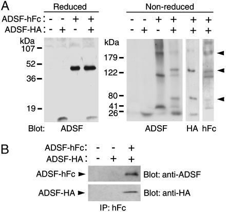
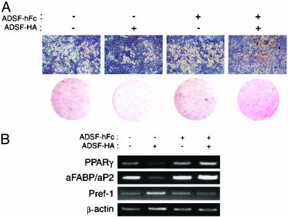
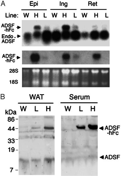
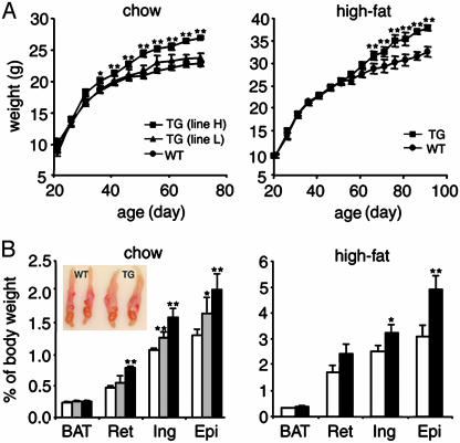
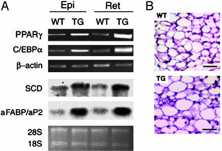
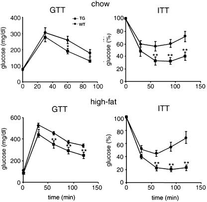
Adipocyte-specific secretory factor (ADSF)/resistin is a small cysteine-rich protein secreted from adipose tissue that belongs to a gene family found in inflammatory zone (FIZZ) or found in resistin-like molecule (RELM). ADSF has been implicated in modulating adipogenesis and insulin resistance. To examine the long-term function of ADSF in adipogenesis and glucose homeostasis, we constructed an expression vector for a dominant inhibitory form of ADSF by fusing it to the human IgGgamma constant region (hFc). ADSF-hFc not only homodimerizes but heterooligomerizes with ADSF/resistin and prevents ADSF/resistin inhibition of adipocyte differentiation of 3T3-L1 cells in a dominant negative manner. Transgenic mice overexpressing ADSF-hFc in adipose tissue show increased adiposity with elevated expression of adipocyte markers as well as enlarged adipocyte size. This finding clearly demonstrates in vivo the inhibitory role of ADSF in adipogenesis. ADSF-hFc transgenic mice with impaired ADSF function exhibit improved glucose tolerance and insulin sensitivity either on chow or high-fat diets. Because of the enhanced adipocyte differentiation, the ADSF-hFc transgenic mice show increased expression of leptin and adiponectin in adipose tissue. The elevated circulating levels for these adipocyte-derived hormones with decreased plasma triglyceride and free fatty acid levels may account for the improved glucose and insulin tolerance in these transgenic mice.


Figures

Fig. 1.
Fig. 1.
Binding of ADSF-hFc to ADSF. 3T3-L1 cells were differentiated in conditioned media from COS7 cells transfected with pcDNA3.1 control vector or ADSF expression vectors. (A) Conditioned media containing ADSF-HA, ADSF-hFc, or both ADSF-HA and ADSF-hFc were subjected to reducing (15%) or nonreducing (6%) SDS/PAGE followed by Western blot analysis with antibodies against ADSF, HA, or human Fc fragment. (B) Conditioned media containing ADSF-HA or ADSF-HA and ADSF-hFc were immunoprecipitated by using anti-hFc agarose beads followed by Western blot analysis with antibodies against ADSF and HA.
Fig. 2.
Fig. 2.
Effect of conditioned media from COS7 cells transfected with ADSFHA, ADSF-hFc, or ADSF-HA and ADSF-hFc on 3T3-L1 adipocyte differentiation. (A) Microscopic pictures for cell morphology and Oil red O staining of 3T3-L1 cells after subjection to differentiation protocol. (B) Adipocyte markers (PPARγ and aFABP/aP2) and a preadipocyte marker (Pref-1) were analyzed by RT-PCR by using total RNA isolated from conditioned-medium-treated 3T3-L1 adipocytes. β-Actin was used as control.
Fig. 3.
Fig. 3.
ADSF-hFc expression in transgenic mice. (A) Northern blot analysis in lines H and L of ADSF-hFc mice and wild-type (W) mice. Total RNA was extracted from the tissues of 10-week-old mice and probed with radiolabeled ADSF (Top) or hFc (Middle) cDNA probes. Epi, epididymal fat pad; Ing, inguinal fat pad; Ret, retroperitoneal fat pad; Endo, endogenous. (B) Western blot analysis for ADSF-hFc fusion protein in serum from lines H and L of ADSF-hFc and wild-type mice. The serum samples were subjected to SDS/PAGE and probed with ADSF antibody. The antibody detected both the 40 kDa of ADSDF-hFc fusion protein and the 12.5 kDa of endogenous ADSF in transgenic mice, but only the 12.5 kDa of ADSF was detected in wild-type mice. Positions of 28S and 18S rRNA are shown.
Fig. 4.
Fig. 4.
Growth curves and fat pad weights of ADSF-hFc transgenic and wild-type mice. (A) Body weight of male mice on a chow or high-fat diet was measured at 5-day intervals; each point represents mean ± SEM; n = 8-9 for each group. (B) Fat pad weights of 10-week-old transgenic mice of line H (filled bar), line L (hatched bar), and wild-type littermates (open bar) are presented as percentage of body weight (n = 8-14 for each group). BAT, brown adipose tissue; Ret, retroperitoneal fat pad; Ing, inguinal fat pad; Epi, epididymal fat pad. Results are mean ± SEM. Statistically significant differences between the groups are indicated: *, P < 0.05. **, P < 0.01. (Inset) Epididymal fat pads from 10-week-old mice of line H and wild-type littermates.
Fig. 5.
Fig. 5.
Adipocyte marker expression and histological analysis of adipose tissue in ADSF-hFc transgenic and wild-type mice. (A) PPARγ, C/EBPα, and β-actin mRNA levels were determined by RT-PCR using total RNA isolated from adipose tissues of 10-week-old ADSF-hFc and wild-type mice fed a chow diet. SCD and aFABP/aP2 were analyzed by Northern blot analysis using 10 μg of total RNA. Epi, epididymal fat pad; Ret, retroperitoneal fat pad. (B) Paraffin-embedded sections of epididymal WAT from 10-week-old mice were stained with hematoxylin and eosin. (Scale bar = 50 μm.) Positions of 28S and 18S rRNA are shown.
Fig. 6.
Fig. 6.
GTTs and ITTs in ADSF-hFc transgenic mice. For GTT, overnight-fasted mice that were fed either a chow or a high-fat diet were given an i.p. injection of glucose (2 mg/g of body weight). Blood samples from the tail were analyzed for glucose concentration. For ITT, mice on chow or high-fat diet were fasted for 5 h before i.p. administration of insulin, 0.5 units/kg of body weight and 0.75 units/kg of body weight, respectively. Glucose (%) represents percent of glucose concentration at 0 min. Each point represents mean ± SEM; n = 6-8 for each group. *, P < 0.05; **, P < 0.01.
Fig. 7.
Fig. 7.
Characterization of mRNA and plasma levels of leptin and adiponectin and fatty acid metabolism in ADSF-hFc transgenic mice. (A) mRNA levels for leptin and adiponectin were measured by RT-PCR using total RNA isolated from epididymal WAT of 10-week-old mice fed a chow diet (Left). β-Actin was used as control. Plasma levels for leptin and adiponectin were measured from overnight-fasted 10-week-old mice fed a chow diet (Right). Results are mean ± SEM from 6-15 mice in each group. Statistically significant differences between the groups are indicated: *, P < 0.05; **, P < 0.01. (B) ACO mRNA levels were determined by Northern blot analysis using total RNA isolated from skeletal muscle of wild-type (lanes 1-3) and ADSF-hFc transgenic mice (lanes 4-6) fed a chow diet. WT, wild-type; TG, transgenic; ACO, acyl-CoA oxidase. Positions of 28S and 18S rRNA are shown.

References

    1. Klyde, B. J. & Hirsch, J. (1979) J. Lipid Res. 20, 705-715. - PubMed
    1. Gregoire, F. M., Smas, C. M. & Sul, H. S. (1998) Physiol. Rev. 78, 783-809. - PubMed
    1. MacDougald, O. A. & Mandrup, S. (2002) Trends Endocrinol. Metab. 13, 5-11. - PubMed
    1. Zhao, L., Gregoire, F. & Sul, H. S. (2000) J. Biol. Chem. 275, 16845-16850. - PubMed
    1. Zhang, Y., Proenca, R., Maffei, M., Barone, M., Leopold, L. & Friedman, J. M. (1994) Nature 372, 425-432. - PubMed

Publication types

LinkOut - more resources